


Crossref Citations
This article has been cited by the following publications. This list is generated based on data provided by Crossref.
Rosli, Siti-Zaharah
Mohd Adzahan, Noranizan
Karim, Roselina
and
Mahmud Ab Rashid, Nor-Khaizura
2022.
Effect of Acidic Electrolysed Water and Pulsed Light Technology on the Sensory, Morphology and Bioactive Compounds of Pennywort (Centella asiatica L.) Leaves.
Molecules,
Vol. 28,
Issue. 1,
p.
311.
Chang, Jia-Dong
Xie, Yun
Zhang, Huanhuan
Zhang, Shurui
and
Zhao, Fang-Jie
2022.
The vacuolar transporter OsNRAMP2 mediates Fe remobilization during germination and affects Cd distribution to rice grain.
Plant and Soil,
Vol. 476,
Issue. 1-2,
p.
79.
Guan, Huize
Jiang, Zhongquan
Sun, Danqing
Wang, Zhongyang
Sun, Yutong
Huo, Hongxun
Li, Zhaoyan
Tang, Lingyi
Li, Zhen
Zhang, Chunhua
and
Ge, Ying
2023.
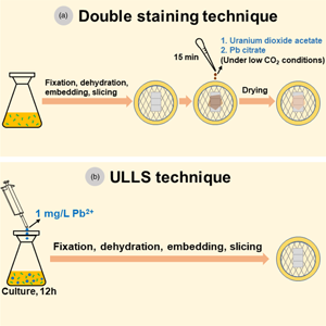
Sufficient Phosphorus Enhances Resistance and Changes Accumulation of Lead in Chlamydomonas reinhardtii.
Environmental Toxicology and Chemistry,
Vol. 42,
Issue. 9,
p.
1960.
Pan, Shang
Li, Zhaoyan
Wang, Jiayi
Li, Xuefei
Meng, Lingzi
Chen, Yunhui
Su, Mu
and
Li, Zhen
2023.
Electron microscopic imaging and NanoSIMS investigation on physiological responses of Aspergillus niger under Pb(II) and Cd(II) stress.
Frontiers in Bioengineering and Biotechnology,
Vol. 10,
Issue. ,