Article

LC-HR/MS Analysis of Lipophilic Extracts from Calendula arvensis (Vaill.) L. Organs: An Unexplored Source in Cosmeceuticals

Department of Environmental, Biological and Pharmaceutical Sciences and Technologies, University of Campania “Luigi Vanvitelli” Via Vivaldi 43, 81100 Caserta, Italy
*
Author to whom correspondence should be addressed.
Molecules 2022, 27(24), 8905; https://doi.org/10.3390/molecules27248905
Submission received: 10 November 2022 / Revised: 7 December 2022 / Accepted: 12 December 2022 / Published: 14 December 2022
(This article belongs to the Special Issue Phytochemistry and Biological Properties of Medicinal Plants)

Abstract

:
As part of a project aimed at promoting the use of Calendula arvensis (Vaill.) L. (field marigold, Asteraceae) phytocomplexes in cosmeceutical formulations, the chemical composition in apolar specialized metabolites is herein elucidated. Furthermore, the screening of the cytotoxicity of the apolar extracts was evaluated in order to underline their safety as functional ingredients for cosmetics. After dissection of Calendula organs (florets, fruits, leaves, bracts, stems, and roots), ultrasound-assisted maceration in n-hexane as an extracting solvent allowed us to obtain oil-like mixtures, whose chemical composition has been highlighted through a UHPLC-ESI-QqTOF-MS/MS approach. Twenty-nine metabolites were tentatively identified; different compounds, among which the well-known poly-unsaturated fatty acids, and oxylipins and phosphatides were detected for the first time in Calendula genus. The screening of the dose-response cytotoxicity of the apolar extracts of C. arvensis highlighted the concentration of 10 μg/mL as the most suitable for the formulation of cosmeceutical preparations. Sera enriched with leaf and fruit apolar extracts turned out to have the best activity, suggesting it can be used as a new source in skin care thanks to their higher content in fatty acids.

1. Introduction

The growing interest of consumers towards ‘everything natural’, coupled with companies’ awareness of an urgent need for a more natural approach to body care, has been the driving force in the replacement of synthetic ingredients, often responsible for some health-associated risks. In this context, wild medicinal and aromatic plants (MAPs) offer unique opportunities of exploitation, as biotic and abiotic factors can influence the content of active molecules [1].
Since ancient times, skin health and appearance improvement have been closely related around the world with the use of natural substances. The interest in natural plant-based products has never waned, and in recent years it has innovated thanks to the growing demand for products that jointly preserve functionality and naturalness [2]. The ancient cosmetic use of plants, and in particular of their oils, to which conditioning, occlusive, soothing, and moisturizing properties are correlated [3,4], is currently being renewed as a precious remedy for the treatment of dermatological disorders, including inflammation, phototoxicity, psoriasis, and atopic dermatitis [5]. In fact, the awareness that oxidative stress plays an important role in skin aging [6], and that prolonged exposure to UV rays reduces the skin antioxidant capacity [7,8] has led to the research and development of multifunctional products based on plant active ingredients. The latter have been extensively investigated due to their ability to protect the skin from exogenous and endogenous factors, and to rebalance lipid homeostasis altered by dermatosis and aging [9]. Thus, natural plant-based cosmetics are proven to promote antioxidant and protective effects, improving skin appearance and preventing UV ray- and oxidative stress-induced damages [10]. In this regard, fatty acids, carotenoids, and polyphenols play an important role. Fatty acids (FAs) are essential for maintaining the normal structure and function of the skin, as they are its constituents. The stratum corneum contains lipids (11%), including water-impermeable glycolipids, adhesive intercellular lipids (cement), and skin coat-forming lipids (NMF) [11], which are responsible for preserving acidic pH and to be an additional protective barrier [12]. Essential fatty acids (EFAs), such as linoleic acid (LA) and α-linolenic acid (ALA), decrease in the epidermis with age, causing sensitivity and roughness of the skin [13], whereas their supplementation, also through topical application, massively improves the skin barrier [14,15]. In addition to fatty acids, carotenoids play a vital role in promoting skin health. Several studies have shown that especially β-carotene, lycopene, lutein, and astaxanthin exert photoprotective effects, due to their light-absorbing properties and their ROS scavenging activity, as well as by regulating UV light-induced gene expression, modulating stress-dependent signaling, and suppressing cellular and tissue responses [16,17,18,19]. Thus, the combination of polyunsaturated fatty acids and carotenoids is to be considered a valid basic strategy for maintaining skin health, and MAPs offer unique opportunities for the recovery and exploitation of these active compounds [1].
In the Mediterranean basin, the Italian vascular flora is the richest in species [20] and represents a precious source of plant resources with potential bioactive molecules poorly studied. In this study, our attention focused on Calendula arvensis (Vaill.) L., known as field marigold, an annual herbaceous Euro-Mediterranean species belonging to the Asteraceae family that recently was deeply investigated for its polyphenol and saponins constituents [21], and new findings are in line with the use of the different organs for diversely recovering antioxidant compounds.
The genus Calendula includes several species known since ancient times for their therapeutic properties [22], even if only Calendula officinalis L. currently has a great interest in the cosmetic market [23,24]. Flower-based preparations of C. arvensis are recommended for external use to maintain skin firmness, to prevent skin inflammation, and to regenerate damaged tissues [25,26], and antioxidant, anticandidal, antifungal, cytotoxic, and antimicrobial activity have been attributed to alcoholic and aqueous extracts [27,28].
Thus, as part of a project aimed at promoting the use of C. arvensis phytocomplexes in cosmeceutical formulations, our interest is turned on deepening the chemical composition of the n-hexane extracts for the valorization of these oil-like mixtures in topical application strategies. Furthermore, this species is very common in its native distribution range and adaptable to different environmental conditions with promising cultivation possibilities.
Therefore, UHPLC-ESI-QqTOF-MS/MS analyses were carried out to evaluate lipophilic constituents in the extracts from the different organs (florets, bracts, fruits, stems, leaves, and roots), whereas after evaluating their dose-response cytotoxicity towards HaCaT and MCF-10 epithelial cell lines, bioactive sera were prepared and their effect on the cellular viability was assessed.

2. Results and Discussion

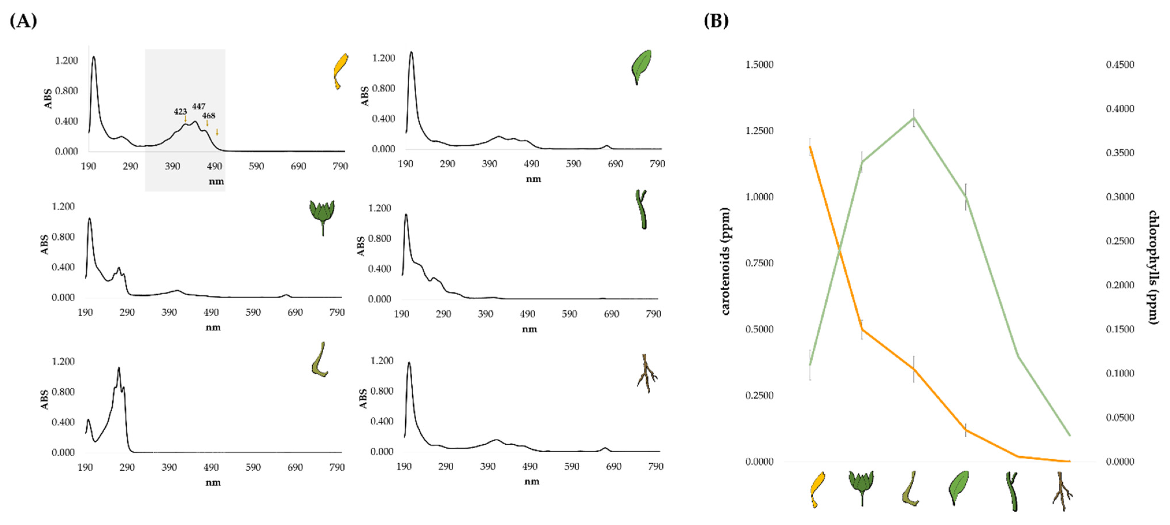
2.1. Lipid Profile of the Different Calendula arvensis Organs

The apolar extracts from florets, bracts, fruits, leaves, stems, and roots of C. arvensis (Figure 1) were preliminarily investigated for their chemical constituents by means of UHPLC-ESI-Qq-TOF-MS/MS analysis.
Based on ultrasound-assisted extraction, and the use of n-hexane as extractant, the diversity in fatty acids of C. arvensis organs was unraveled, and twenty-nine metabolites were tentatively identified (Table 1, Table 2, Table 3 and Table 4). Indeed, fatty acids (FAs) were mainly in their free form, whereas glycerol-phosphorylated FAs were detected as minor constituents, together with triterpene aglycones and glycosides. In Table 1, TOF-MS and TOF-MS/MS data of tentatively identified fatty acids are reported.
The most abundant compounds were α-linolenic acid (15) and its constitutional isomers (16, 17), as well as linoleic acid (19). They were previously identified in leaves, seeds, and flowers of a petroleum ether extract of C. officinalis L. [29,30]. In particular, the TOF-MS spectra of compounds 15, 16, and 17 showed the [M-H] ion at m/z 277.2177, according to the molecular formula C18H30O2 (Figure 2). Calendic acid isomers (16 and 17), mainly present in fruit organs, were discriminated by α-linolenic acid (15) by means of UV-DAD spectra. In fact, due to the occurrence of three conjugated double bonds, their characteristic λmax at 262, 270, and 282 nm were observed [31,32]. These conjugated trienoic acids were likely α-, and β-calendic acids, whose biosynthesis was from linoleic acid by means of a (1,4)-desaturase [33].
Linoleic acid deprotonated molecular ion (19) was at m/z 279.2336, whereas its isomer, likely the linoelaidicic acid (22) [30], was also distinguished in florets, leaves, and stems extracts. The [M-H] ion at m/z 281.2490 (28) was in accordance with the monounsaturated oleic acid, whereas the deprotonated ions at m/z 253.2177 (18) was likely palmitoleic acid. Among saturated FAs, palmitic acid (24) with the deprotonated molecular ion at m/z 255.2331, and stearic acid (29; [M-H] ion at m/z 283.2643) were tentatively identified in all the investigated extracts. These FAs were previously found as constituents of C. arvensis flowers ethyl acetate extract, the latter showing a promising anti-cancer activity against breast cancer cell lines [28]. Based on TOF-MS/MS spectra in negative ion mode, FAs differently shared the [M-H-44] and [M-H-18] fragment ions [34,35]. Oxygenated FAs also occurred, and favorably fragmented through β-scission and ene reactions (Table 2). Compounds 5 and 10 with ion [M-H] at m/z 295.2284(0), sharing the molecular formula C18H32O3, were tentatively identified as hydroxyoctadecadienoic acids. The different fragmentation pattern of the two compounds allowed us to localize the hydroxyl group at C-13 and C-9, respectively. In particular, the 13-hydroxy derivative (5) showed abundant product ions at m/z 195.1395, which can result from both charge-driven and charge-remote (ene reaction) allylic fragmentation after [1,5]-H sigmatropic shift of the Δ9,11 double bonds [36]. The 9-hydroxy derivative was previously found in C. officinalis seeds oil [31], with TOF-MS/MS spectra providing the characteristic fragment ion at m/z 171.1026, due to oxononanoate [37]. An unsaturated keto FA with [M-H] ion at m/z 291.1968 (3) was tentatively identified as 13-oxo-octadeca-9,11,15-trieonic acid, whereas compounds 7 and 9, sharing the deprotonated molecular ion at m/z 293.2122, were likely oxo-octadecadienoic acids, whose recent detection was also in the fruits extract of C. aegyptaica Desf [38]. Several mechanisms have been proposed for the biosynthesis of these fatty acids, including formation of an epoxy derivative of linoleic acid as intermediate, oxidation or isomerization of linolenic acid, and formation of a linoleic acid radical by a lipoxygenase-type reaction [32]. Indeed, most of the minor compounds in the investigated extracts were phosphatidic acids (PAs; Table 3). In particular, compounds 1, 4, 6, 8, and 12 shared the monoacyl glycerol phosphate core, whereas compounds 20, 23, and 2527 were diacyl derivatives.
Compounds 1 and 4 with [M-H] at m/z 431.2208(07) and molecular formula C21H37O7P, were tentatively identified as isomers of linolenoyl glycerol phosphate (Figure S1A,B). The neutral loss of 154.00 Da was diagnostic of glycerol phosphate, and provided the fragment ion at m/z 277.2172(71). Furthermore, the ion at m/z 96.96 was in accordance with phosphate residue. Analogously, compounds 6 and 8 with [M-H] ion at m/z 433.2364(8) were linoleoyl glycerol phosphate isomers (Figure S1C,D), and compound 12 with [M-H] ion at m/z 409.2366 was likely palmitoyl glycerol phosphate (Figure S1E). The identity of the acyl moiety was through the detection of the ion at m/z 279.2308(6), corresponding to linoleate, in the TOF-MS/MS spectra of the first two compounds, and of the ion at m/z 255.2337 (deprotonated palmitic acid) in the spectrum of compound 12.
The main TOF-MS/MS fragment ions from the diacyl glycerol phosphates (20, 23, and 2527) arose from the neutral loss of acyl moieties as free fatty acids, and/or of FA-glycerol phosphate moiety (Figure S2). When the monoacyl-glycerol phosphate ion was formed, the nucleophilic attack on the C-1 or the C-2 carbon of the glycerol provided [R1CO2] and [R2CO2] ions by charge transfer. Thus, compound 20 was tentatively dilinolenoylglycerol phosphate, so much so that its deprotonated molecular ion at m/z 691.4349 underwent the neutral loss of linolenic acid (−278 Da) to achieve the fragment ion at m/z 413.2115, which in turn lost an allyl hydrogen phosphate to provide the base peak at m/z 277.2174 (linolenate). Glycerol phosphate was detected at m/z 152.9958. Compounds 23 and 25 were likely linolenoyl-palmitoyl glycerol phosphate. The [M-H] ion at m/z 671.4672 (26) lost 280 Da forming the fragment ion at m/z 391.2271, likely corresponding to (palmitoyloxy) allyl hydrogen phosphate, or, alternatively, the neutral loss of linoleoyl moiety as a ketene was observed. The ions at m/z 279.2333 and 255.2335 were in accordance with linoleate and palmitate, respectively. Finally, in compound 27 the phosphatidyl acyl groups are both linoleoyl. Compounds 11 and 21 were tentatively identified as isomers of 16:0/18:2-phosphatidylinositol (Figure S3). The fragmentation pattern resembled that of the glycerol phosphate derivatives described above, and the neutral loss of 162.05 Da was related to inositol moiety. Thus, the deprotonated molecular ion underwent octadecadienoic acid loss to supply the ion at m/z 553.2815(07), which in turn, after losing dehydrated inositol, generated the palmitoyl-dehydroglycerol phosphate moiety (m/z 391.2259(71)). Although these metabolites were not previously isolated in the Calendula genus, they were recently found in fruits of Kigelia africana Benth [39], in seeds of Camelina sativa Crantz, and in Hordeum vulgare L. roots [40]. Recently, phosphatidic acid was reported to be involved in several cell functions in plants, animals, and microorganisms as a lipid messenger [41]. PAs are associated with various regulatory processes, such as signaling pathways in cell growth, proliferation, and reproduction, as well as responses to hormones and biotic and abiotic stresses [42]. Furthermore, it was shown to exert an antiapoptotic effect on epidermal keratinocytes exposed to oxidative stress caused by hydrogen peroxide [43].
Finally, compound 2, 13, and 14 was tentatively identified as triterpenes (Table 4). In particular, compounds 13 and 14 showed the deprotonated molecular ion at m/z 455.3536(7), and were identified as ursolic acid and oleanolic acid, respectively. GC-MS analysis previously noticed the presence of these compounds in the flower extract of C. officinalis L., and literature data showed their ability to exert anti-inflammatory [44], neuroprotective [45], and antimicrobial [46] activities. Compound 2 was tentatively identified as the acetyl derivative of oleanolic acid glucuronide (Figure S4). In fact, in the TOF-MS/MS spectrum the deprotonated molecular ion at m/z 673.3998 provided the fragment ion at m/z 631.3903 through the loss of 42 Da (-COCH3), which in turn likely underwent water loss to achieve the ion at m/z 613.3746. Furthermore, the hexuronyl moiety loss (−176 Da) was displayed by the deprotonated molecular ion. The loss of 62 Da from the ion at m/z 673.3998 to provide the ion at m/z 569.3872 further confirmed the occurrence of hexuronyl unit [21]. This neutral loss was previously ascribed to simultaneous decarboxylation and dehydration of the saccharidic part [21]. Indeed, C. arvensis has been described as source of triterpene saponins based on oleanolic and echinocystic acid. The diversity in the knowledge of these compounds in the Calendula genus is herein enhanced by the identification of the compound 2.
The relative quantitation of the tentatively identified fatty acids, oxylipins, phosphatides, and triterpenes highlighted that free fatty acids were the most abundant compounds in all the organs, with the highest amount in leaves and involucral bracts. Principal component analysis (PCA) was carried out considering the relative content of each identified free fatty acid (Figure 3). The two principal components (PC1 and PC2) account for 98.03% of the total variance; the first principal component represents the 90.13% with linoleic acid as the most abundant FAs in all the extracts, being located at the end of the positive score. PC2 underlined leaf and stem richness in calendic acid (1), whereas calendic acid (2) exceptionally was in fruit extract.
The heatmap of the apolar extracts from C. arvensis organs showed that cluster segregations occurred based on the relative composition of each investigated organ (Figure 4). In particular, this proves that florets mainly differed for their triterpene content, while leaves were rich in FAs, and seeds in OxFAs. Finally, phospatides were more abundant in stems and leaves than in the other organs.
Furthermore, to have a clear picture without deepening the carotenoid compositional detail, the photosynthetic pigments’ content among the different organs was unraveled through UV analysis. The acquired UV-Vis spectra of all the investigated organs (Figure 5A) showed that florets contained an appreciable amount of carotenoid compounds, which are poorly represented also in leaves, stems, and involucral bracts. Florets richness in carotenoids was inversely related to their low chlorophyll content (Figure 5B). Indeed, the UV-Vis spectrum of involucral bracts also detected calendic acid, which further dominated in fruits. According to literature, large amounts of carotenoids have been found in the flowers of the more common C. officinalis but with a wide variety of contents, ranging from little to large quantities [47,48,49], which are related to several factors such as plant variety, color of the ligulate and tubular florets, site of cultivation, and harvesting time [47,50,51,52,53].
In the petals of C. officinalis, the main carotenoids found were flavoxanthin and auroxanthin [50], while the stems and leaves mostly contained lutein and carotene [48,54]. Carotenoids are biologically active compounds broadly applied in cosmetics, they can act as free radical scavengers and can protect healing wounds [55]. Lutein and its esters are the most abundant carotenoids. This xanthophyll is the basis of supplements useful to reduce the risk of developing age-related macular degeneration (AMD), a degenerative disease, which causes irreversible blindness in the elderly [56]. Only recently an exhaustive investigation was launched on carotenoids and it is necessary to deepen the knowledge of the content of the florets in C. arvensis.

2.2. Cytotoxicity Screening of Apolar Extracts from Calendula arvensis Organs

HaCaT human keratinocytes and MCF-10 non-tumorigenic epithelial cell lines were used for carrying out the in vitro evaluation of the C. arvensis organs’ cytotoxicity through MTT test. HaCaT cells form a reliable in vitro model for studying the functions of keratinocytes, which constitute the 95% of the epidermal cells, as inflammatory/repair response [57] or wound healing effects of plant extracts [58]. MCF-10 cell lines, with a structural similarity to the normal human mammary epithelium, are widely used in cytotoxicity studies as a control to evaluate the safety of different compounds in biomedicine [59,60] and personal care products [61,62]. Thus, both cell lines represent valid models for cytotoxicity tests in dermoprotection studies. After treating cells with increasing doses of C. arvensis apolar extracts, the obtained data, analyzed as the average of three replicates, were organized in a matrix (6 organs × 5 concentrations) for each of the two used cell lines. Successively, each data matrix was processed by cluster analysis to explore the degree of values between plant organs and concentration tested. An average linkage agglomeration criterion and Jaccard Index as dissimilarity coefficient were applied.
The obtained dendrograms (Figure 6) clearly showed different clustering patterns of the organs apolar extracts, depending on the cell lines used.
In particular, dendrogram referred to an HaCaT cell line that evidenced two main clusters with a dissimilarity value of 50% (Figure 6A). The first cluster included only the fruit extract, while the second consisted of three subclusters which in turn contained: roots, florets, leaves, bracts, and stems. Cluster analysis on MCF-10 cell line also revealed two main clusters but with 80% of dissimilarity value and each, in turn, composed of two subclusters. The first cluster consisted of the subcluster of leaves, and that grouping florets and bracts. The second cluster also distinguished two subgroups: one containing only stems, and the other with roots and fruits (Figure 6B).
Afterwards, the organ activity data were ordered according to the obtained groups of each dendrogram, respectively (Figure 6C,D), to highlight the cytotoxicity trends in relation to the tested extract concentrations.
With regards to HaCaT cell line, it is clearly observed that the subcluster florets and leaves, as well as that of stems and bracts, are related to the less cytotoxic organs, followed by florets and leaves. All these organs showed a similar behavior in terms of cytotoxicity increase while increasing the concentration. The roots, on the other hand, exhibited a maximum of cytotoxicity at a concentration of 50 μg/mL, whereas the fruit extract showed a very different behavior compared with the other organs, highlighting a strong activity already at the 10 μg/mL tested dose. The data obtained appeared in line with the chemical composition of the organs analyzed. In fact, considering the diversity of florets in triterpenes and phosphatidic acid, it is known that triterpenes exert anti-inflammatory [63], healing [64], and antiproliferative [65] activities. In particular, Ghosh et al. [66] showed that oleanolic acid induced non-appreciable cell death in human keratinocytes compared with cancer cell line, and ursolic acid was involved in reducing the release of pro-inflammatory cytokines NF-κB, IL-6, and TNF-α [67]. On the other hand, phosphatidic acids, which play a key role in intracellular signaling, have been shown to directly activate pro-inflammatory protein kinases [68], and are a source of glycerol and fatty acids. Glycerol exerts beneficial effects on the epidermis by improving the hydration of the stratum corneum, the skin barrier function, and the mechanical properties of the skin, the inhibition of the lipid phase transition of the stratum corneum, the protection from irritating stimuli, the improvement of degradation desmosomal and acceleration of wound healing processes [69]. Furthermore, implementation with n-3 and n-6 PUFAs showed improvement in psoriasis and atopic dermatitis [70]. Recent evidence showed that PUFAs belonging to n-3 class improve inflammatory skin, and the exogenous supplementation of α-linolenic was observed to enhance the skin barrier function thanks to its ability to be incorporated into the phospholipid and triglyceride fractions of the skin. Thus, it can act as modulator of lipid mediators, such as prostaglandins, hydroxy fatty acids, and monoacylglycerols [15].
Oxylipins, which are important constituents in the stem, root, and fruit extracts, are pleiotropic modulators of metabolic and inflammatory responses [71], being regulators differently during cell proliferation, differentiation, and migration [72]. Thus, their higher concentration in the root and fruit extract can explain the higher cytotoxic effect observed. Indeed, in a complex entity such as a plant extract, whose chemistry strongly depends on the extraction method applied and plant organ intrinsic chemical composition [73], the effect can be present through multiple mechanisms. Furthermore, a time- and dose-dependent cytotoxicity can occur, so much so that extracts not exhibiting cytotoxic effects at lower doses are pro-oxidant and cytotoxic when tested at higher doses. Although there are no data in the literature about the relationship between phytochemicals from C. arvensis and cytotoxicity profile, the controversial role of C. officinalis extracts as a topical agent, also able to be involved in radiodermatitis treatment, was reviewed [74], and extracts obtained through distillation or percolation referred to contain phenolic compounds based on Folin–Ciocalteau assay, were observed as protective in HaCaT cells subjected to hydrogen peroxide insult [75]. Analogously, an aqueous extract from C. officinalis was observed to protect HaCaT cells against detrimental effects of oxidative stress-inducing personal care products [76]. The precious diversity in essential fatty acids, and their derivatives in investigated extracts suggested their potential use for counteracting changes in skin lipid components, which can contribute to the onset of different skin diseases. Thus, based on this preliminary cytotoxic screening, the most suitable concentration of the organ extracts to be used in the subsequent phase of the formulation with serum addition was established. In fact, excluding the 50 μg/mL concentration of all the organs, which was highly cytotoxic towards both the tested cell lines, the concentration of 10 μg/mL was chosen as the maximum concentration with the lowest toxicity suitable for formulating cosmeceutical preparations. Although for the chosen concentration, inhibitions of mitochondrial redox activity were observed by the seed extract on HaCaT cells and by the stem extract on the MCF-10 cell line, the concentration was nevertheless adopted for the inclusion of the extract in the formulation.

2.3. Cytotoxicity of Cosmeceutical Formulations Based on Calendula arvensis Apolar Extracts

MTT data on HaCaT and MCF-10 cell lines put the basis for the use of the different organs apolar extracts in the development of a cosmeceutical preparation, which can guarantee a beneficial effect on skin. The serum is a highly concentrated product with properties of rapid absorption and the ability to penetrate through the phospholipid bilayer cells. It provides ten times greater richness of biologically active substances than O/W emulsions, thus performing faster and more successfully for skin-related problems [77]. Therefore, the prepared acqueous sera, each one enriched with a different extract (0.2% p/v), underwent an in vitro screening in order to highlight their cytotoxic potential.
The principal component analysis of %RAI of the serum-based apolar extracts makes clear the cytotoxicity gradient along the first axis (PC 1) related to the tested organs and cell lines, with HaCaT being less cytotoxic than MCF-10, and the latter in turn less cytotoxic of the aqueous serum used as blank (Figure 7A). In particular, it was observed that sera enriched with apolar extracts from roots, leaves, and fruits exhibited a similar behavior in both cell lines. Conversely, bracts, flowers, and stems produced greater dissimilarity effects in relation to the target cell line and, in general, a lower cytotoxicity was observed on HaCaT compared with MCF-10.
The RAI percentage of each analyzed organ ordered along axis 1 as depicted by PCA %RAI of each organ showed a low cytotoxic activity (Figure 7B) that does not exceed the value of 20%. In fact, the high inhibition %, equal to 14.07 ± 0.52 was recorded by stem extract, which alone appeared to be more cytotoxic. This trend was in agreement with Balestrin et al. [78] in which the incorporation of Achyrocline satureioides (Lam.) DC ethanolic extract into nanoemulsion formulation reduce the cytotoxicity on HaCaT cells compared with free extract treatment [79].
Among all organ extracts-enriched sera tested (Figure 7B), leaves and fruits-included sera showed the best activity with RAI percentage values ranging from very low (0.49 + 0.39 in leaves and 0.31 + 0.53 in fruits) on HaCaT cells, to low (3.32 + 0.98 in leaves and 3.19 + 0.42 in fruits) on MCF-10 cells, thus suggesting that the enriched sera can be considered available and safe thanks to their content in fatty acids and their derivatives. Conversely, flowers and stems displayed a low cytotoxic activity on HaCaT cells, or proliferative activity on MCF-10. This cell-sensitive effect can be due to their triterpene content, which was only partially mitigated by the serum. In fact, previous data underline that ursolic acid was able to induce apoptosis through caspase-3 activation and cell cycle arrest in HaCat cells [80].
To date, there is extensive knowledge of the cosmeceutical preparation of C. officinalis, which has 14 INCI designations in the European list of cosmetic ingredients [81]. An aqueous formulation with bio-soothing functional compounds of C. officinalis is commercially available, as well as various other C. officinalis preparations, mainly extracts, tinctures, and oils to be incorporated into topical formulations aimed at wound healing and to soothe inflamed and damaged skin. C. officinalis flower extract is the most used in cosmetic products [82]. Our results on C. arvensis are the first to highlight and suggest a potential exploitation of some organs of this species as a new source for skin care, with indications on extract concentration to be used.
As mentioned above, lipids play a crucial role in maintaining normal skin function, and topical application of a formulation enriched in fatty acids extracts can promote this process. Furthermore, free PUFAs have important regulatory effects on the initiation, development, and resolution of inflammation as they can be converted into hundreds of lipid-modulating substances [83]. In this scenario, considering the close link between chemical composition and biological activity, leaves- and fruits-enriched sera represent the most valuable resource for fatty acids topical application being rich in this class of compounds.

3. Materials and Methods

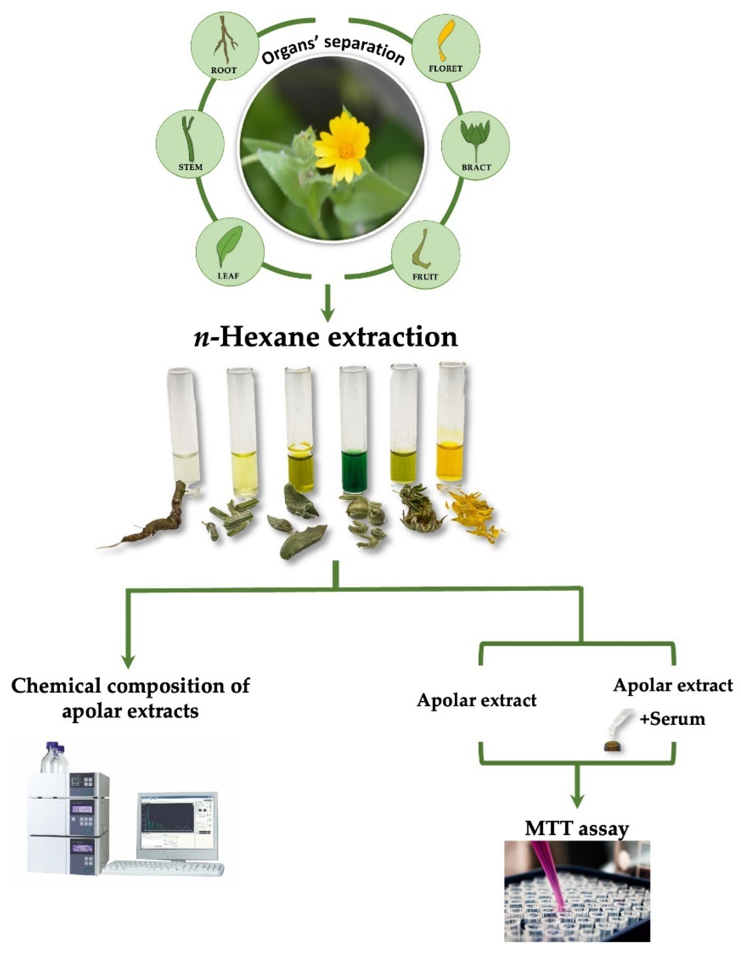
3.1. Plant Collection and Extraction

Calendula arvensis (Vaill) L. was harvested in May 2021 in Roccaromana (latitude 4°16′24.8″ N, longitude 14°12′56.9″ E; 163 m a.s.l), southern Italy. The environmental site characteristics, data on the voucher specimen and organs separation procedure are reported by Fiorentino et al. [21].
The different organs florets, bracts, fruits, stems, leaves, and roots obtained from plant dissection, have been freeze-dried for 3 days using the FTS System Flex-DryTM instrument (SP Scientific, Stone Ridge, NY, USA). The different cryo-dried organs were pulverized by a rotary knife homogenizer (Knife Mill PULVERISETTE 11, Buch & Holm, Herlev, Denmark) and a sample (∼5.0 g) underwent solid–liquid extraction by ultrasound-assisted maceration (UAM; Branson UltrasonicsTM BransonicTM M3800-E, Danbury, CT, USA) using n-hexane as an extracting solvent. Three extraction cycles (30 min each) were performed and at the end of each cycle the sample was filtrated and the extraction solvent was removed using a rotary evaporator (Heidolph Hei-VAP Advanyage, Schwabach, Germany). The apolar extract of each organ was chemically analyzed by UHPLC-ESI-QqTOF-MS/MS (Shimadzu, Tokyo, Japan; AB Sciex, Concord, ON, Canada) and HPLC-UV-DAD analyses (Agilent, Santa Clara, CA, USA). Moreover, the cytotoxicity was assessed on both organs’ extracts and the sera enriched with them (Figure 8).

3.2. UHPLC-ESI-QqTOF-MS/MS Analyses

The apolar extracts from the different C. arvensis organs were investigated using a NEXERA UHPLC system (Shimadzu, Tokyo, Japan) equipped with a Luna® Omega C-18 column (50 × 2.1 mm i.d., 1.6 μm particle size). The mobile phase was constituted by water (solvent A) and acetonitrile (solvent B), both acidified with formic acid (0.1% v/v). A linear gradient was used, in which the percentage of solvent B increased as follows: 0–12 min, 5%→32% B; 12–30 min, 32%→75% B; 30–31 min, 75%→95% B; 31–32 min, 95% B. The mobile phase composition was allowed to re-equilibrate for 2 min. The flow rate was set at 0.5 mL/min. High-Resolution Mass Spectrometry (HR-MS) data were obtained by an AB SCIEX Triple TOF® 4600 mass spectrometer (AB Sciex, Concord, ON, Canada), equipped with a DuoSprayTM ion source (AB Sciex, Concord, ON, Canada) operating in the negative ElectroSpray (ESI) mode. A full scan Time-of-Flight (TOF) survey (accumulation time 100 ms, 100–1000 Da) and 8 information-dependent acquisition MS/MS scans (accumulation time 50 ms, 80–850 Da) were acquired using the following parameters: curtain gas 35 psi, nebulizer and heated gases 60 psi, ion spray voltage 4500 V, ion source temperature 600 °C, declustering potential −80 V, and collision energy −40 ± 15 V. The instrument was controlled by Analyst® TF 1.7 software (AB Sciex, Concord, ON, Canada), whereas MS data were processed by PeakView® software version 2.2 (AB Sciex, Concord, ON, Canada). The compounds were identified mainly through the study of their tandem mass spectrometry (TOF-MS/MS; AB Sciex, Concord, ON, Canada) fragmentation patterns, and the comparison with literature data whenever possible.

3.3. HPLC-UV-DAD Analyses

To achieve UV-DAD information of fatty acids isomers, separation was also performed by using a 1260 Infinity II LC System (Agilent, Santa Clara, CA, USA) equipped with an Agilent G711A quaternary pump and a WR G7115A diode array detector. The instrument was equipped with Kinetex® PS C-18 (50 × 2.1 mm i.d., 2.6 μm particle size) with a linear gradient in which the percentage of B increases as the following: 0–5 min, 5%→55% B; 5–10 min, 55%→75% B; 10–11% min, 75%→95% B; 11–12 min, and 95% B. The wavelengths were 205, 268, and 282 nm. The flow was 0.4 mL/min.

3.4. Chlorophyll and Carotenoids Content

The chlorophyll and carotenoids content of C. arvensis n-hexane extracts was spectrophotometrically measured by a Cary 100 Spectrophotometer (Agilent, Santa Clara, CA, USA) against a blank [84]. The chlorophyll and carotenoid content were calculated according to Aladić et al. [85].

3.5. Aqueous Serum Formulation

The aqueous serum (100 g) was prepared adding distillated water (70 g) and glycerol (28.5 g) in a glass beaker. The mixture was stirred until the solution was homogenous. Then sodium alginate (1.5 g), used as swelling agent, was added and the solution was mixed by a homogenizer immersion mixer. The mixture was left to rest until it was free of any lamps. All apolar extracts, previously solubilized in pure ethanol, were incorporated in aqueous serum at 0.2% p/w. The sera, being water-based, are suggested to have a light and rapidly absorbed consistency to penetrate as much as possible into the skin.

3.6. Cell Culture and Cytotoxicity Assessment

Human primary keratinocytes cell lines (HaCaT) were cultured in Dulbecco’s Modified Eagle’s Medium (DMEM) supplemented with 10% fetal bovine serum, 50.0 U/mL of penicillin and 100.0 μg/mL of streptomycin, at 37 °C in a humidified atmosphere containing 5% CO2. Non-tumorigenic epithelial cell lines (MCF-10) were grown in the same conditions except for the medium, which was FBS-free medium supplemented with 10% Horse Serum.
Cells were seeded in 96-multiwell plates at a density of 1.5 × 10⁴ cells/well. After 24 h, cells were treated with five concentrations of the different organs’ apolar extracts (1.0, 2.5, 5.0, 10, and 50 μg/mL). After 5 h of incubation, cells were treated with 20 mg of MTT (3-(4,5-dimethyl-2-thiazolyl)-2,5-diphenyl-2H-tetrazolium; 0.5 mg/mL), dissolved in the FBS-free culture medium, and allowed to stand for 4 h at 37 °C in a 5% CO2 humidified atmosphere. The MTT solution was then removed and 500.0 μL of DMSO were added to dissolve the produced formazan dye. Finally, the absorbance at 570 nm of each well was determined using a Victor3 Perkin Elmer absorbance reader (Perkin Elmer/Wallac, Waltham, MA, USA). Cell viability was expressed as a percentage of mitochondrial redox activity (RAI, %) of the cells treated with the extracts compared with the untreated control, using the following formula [86]:
[(Abs untreated cells) − (Abs treated cells)/(Abs untreated cells)] × 100
Both cell lines seeded in 6-multiwell plates to evaluate the cytotoxicity of serum, used as blank, and sera enriched with the different apolar extracts. The density of HaCaT was 5.0 × 10⁵ cells/well whereas, for MCF-10 was 5.0 × 10⁵ cells/well. The serum concentration used as blank was 36 mg/mL and the formulation enriched was 36 mg/mL plus 1 mg/mL of the apolar extract. After 24 h cells were treated with serum (blank) and serum with all extracts. After 5 h of incubation, MTT (3-(4,5-dimethyl-2-thiazolyl)-2,5-diphenyl-2H-tetrazolium; 0.5 mg/mL) cells’ viability test was carried out as previously described.

3.7. Statistical Analysis

A multivariate analysis by ClustVis (https://biit.cs.ut.ee/clustvis/) [87] was applied to explore and clarify quali-quantitative compositive data compounds in each organ. Numerical clustering of MTT assay data was performed to explore the degree of dissimilarity values between plant organs and concentrations tested by using the SYN-TAX software (SYN-TAX 2000, Syntax, Berlin, Germany) [88].

4. Conclusions

With the prominent role that fatty acids have in epidermal and metabolic pathways, their analysis is of major interest for the formulation of best products in cosmeceutical research field. The apolar extract obtained with different techniques are usually profiled by means of gas chromatography mass spectrometry (GC-MS). Instead, our work demonstrated that liquid chromatography (LC-MS) represents a pivotal tool for determining the chemical profile of oil-like mixtures, focusing on their EFAs composition. In fact, in C. arvensis different classes of compounds not only the well-known PUFAs, but also oxylipins and phosphatides were detected for the first time, widely improving the knowledge of the fatty acid composition of each specific organ of this species and of the whole Calendula genus (Table 5). Furthermore, the biological value of FAs confirms that C. arvensis offers greater resources to apply in the formulation of a high-functional cosmetic. Specifically, the different organs studied underline that each part of the plant represents a reservoir of specific class of compounds, as PUFAs were the most abundant in leaf and fruit extracts. Lipids in cosmetics are designed to be applied to human skin to preserve and improve its appearance, to form a protective barrier, to preserve from external harmful substances and to promote hydration. PUFAs are commonly used as the main ingredient of personal care products, and the increasing demand of natural-base formulation encourages the deeper investigation of medicinal and aromatic plants (MAPs) as both a traditional and innovative source of bioactive compounds. In this regard, C. arvensis proved to be a good candidate to be explored in this field due to its richness and diversity in all the parts of the plant, in different lipophilic compounds clarified by UHPLC-QqTOF- MS/MS analysis. The preliminary cytotoxic screening on two different cells highlighted that the prepared cosmetic, enriched products are safe. Unlike most of the literature data that focused on aerial parts of Calendula genus, this work put the basis for intensifying the study of the relationship between chemical composition and bioactivity (wound healing and anti-inflammatory) of each organ that can be applied for specific therapeutic purposes.

Supplementary Materials

The following supporting information can be downloaded at: https://www.mdpi.com/article/10.3390/molecules27248905/s1, Figure S1: TOF-MS/MS spectrum of the compounds (A) 1, (B) 4, (C) 6, (D) 8, (E) 12 and proposed fragmentation pathway of monoacyl-PAs tentatively identified in apolar extracts from the different C. arvensis organs; Figure S2: TOF-MS/MS spectrum of the compounds (A) 20, (B) 25, (C) 27, and (D) 26, and proposed fragmentation pathway of diacyl-PAs tentatively identified in apolar extracts from the different C. arvensis organs; Figure S3: TOF-MS/MS spectrum of the theoretical [M-H] ion for compound 11.

Author Contributions

Conceptualization, S.P. (Severina Pacifico) and A.E.; methodology, S.P. (Severina Pacifico) and A.E.; formal analysis, C.G., M.F. (Marialuisa Formato), M.F. (Marika Fiorentino) and M.T.P.; investigation, C.G., M.F. (Marialuisa Formato), M.F. (Marika Fiorentino), M.T.P. and S.P. (Simona Piccolella); resources, A.S., S.P. (Severina Pacifico) and A.E.; data curation, C.G., S.P. (Severina Pacifico) and A.E.; writing—original draft preparation, C.G., S.P. (Severina Pacifico) and A.E.; writing—review and editing, C.G., S.P. (Severina Pacifico) and A.E.; visualization, M.F. (Marialuisa Formato), M.F. (Marika Fiorentino), S.P. (Simona Piccolella) and A.S.; supervision, S.P. (Severina Pacifico) and A.E. All authors have read and agreed to the published version of the manuscript.

Funding

This research received no external funding.

Institutional Review Board Statement

Not applicable.

Informed Consent Statement

Not applicable.

Data Availability Statement

Not applicable.

Acknowledgments

The authors thank the PON Project “Valorizzazione delle specie officinali della flora autoctona del Parco Nazionale del Cilento, Vallo di Diano e Alburni (PNCVDA)” DOT13499551.

Conflicts of Interest

The authors declare no conflict of interest.

Sample Availability

Samples of the field marigold oil-like mixtures are available from the authors.

References

  1. Piccolella, S.; Crescente, G.; Pacifico, F.; Pacifico, S. Wild aromatic plants bioactivity: A function of their (poly)phenol seasonality? A case study from Mediterranean area. Phytochem. Rev. 2018, 17, 785–799. [Google Scholar] [CrossRef]
  2. Alcalde, M.T. Natural and ecological cosmetics. Offarm 2008, 27, 96–104. (In Spanish) [Google Scholar]
  3. Burnett, C.L.; Fiume, M.M.; Bergfeld, W.F.; Belsito, D.V.; Hill, R.A.; Klaassen, C.D.; Liebler, D.; Marks, J.G., Jr.; Shank, R.C.; Slaga, T.J.; et al. Safety Assessment of Plant-Derived Fatty Acid Oils. Int. J. Toxicol. 2017, 36, 51S–129S. [Google Scholar] [CrossRef] [PubMed]
  4. Sánchez-Vioque, R.; Herraiz-Peñalver, D.; Melero Bravo, E.; Ortiz de Elguea-Culebras, G.; Herrero, B.; Santiago, Y.; Bueno, M.; Pérez-Magarino, S.; del Carmen Asensio, S.; Manzanera, M. Variability of the essential oil composition of cultivated populations of Salvia lavandulifolia Vahl. Crop Sci. 2022, 62, 744–752. [Google Scholar] [CrossRef]
  5. Aburjai, T.; Natsheh, F.M. Plant Used in Cosmetics. Phytother. Res. 2003, 17, 987–1000. [Google Scholar] [CrossRef]
  6. Gilchrest, B.A.; Krutmann, J. Skin Aging, 1st ed.; Springer: Berlin/Heidelberg, Germany, 2006; pp. 33–42. [Google Scholar]
  7. Bouftira, I.; Abdelly, C.; Sfar, S. Characterization of cosmetic cream with Mesembryanthemum crystallinum plant extract: Influence of formulation composition on physical stability and antioxidant activity. Int. J. Cosmet. Sci. 2008, 30, 443–452. [Google Scholar] [CrossRef]
  8. Auner, B.G.; Wirth, M.; Valenta, C. Antioxidative activity and cytotoxicity of four different flavonoids for dermal applications. J. Drug Deliv. Sci. Technol. 2005, 15, 227–232. [Google Scholar] [CrossRef]
  9. Siti Zulaikha, R.; Sharifah Norkhadijah, S.I.; Praveena, S.M. Hazardous Ingredients in Cosmetics and Personal Care Products and Health Concern: A Review. J. Public Health Res. 2015, 5, 7–15. [Google Scholar] [CrossRef]
  10. Draelos, Z.D. The Multifunctional Value of Sunscreen containing Cosmetics. Skin Ther. Lett. 2011, 16, 1–3. [Google Scholar]
  11. Spector, A.A.; Yorek, M.A. Membrane lipid composition and cellular function. J. Lipid Res. 1985, 26, 1015–1035. [Google Scholar] [CrossRef]
  12. Nicolaou, A. Eicosanoids in skin inflammation. Prostaglandins Leukot. Essent. Fat. Acids 2013, 88, 131–138. [Google Scholar] [CrossRef] [PubMed]
  13. Wertz, P.W. Lipids and barrier function of the skin. Acta Derm. Venereol. Suppl. 2000, 208, 7–11. [Google Scholar] [CrossRef] [PubMed]
  14. Bialek, A.; Bialek, M.; Jelinska, M.; Tokarz, A. Fatty acid profile of new promising unconventional plant oils for cosmetic use. Int. J. Cosmet. Sci. 2016, 38, 382–388. [Google Scholar] [CrossRef] [PubMed]
  15. Simard, M.; Tremblay, A.; Morin, S.; Martin, C.; Julien, P.; Fardette, J.; Flamand, N.; Pouliot, R. α-Linolenic acid and linoleic acid modulate the lipidome and the skin barrier of a tissue-engineered skin model. Acta Biomater. 2022, 140, 261–274. [Google Scholar] [CrossRef]
  16. Stahl, W.; Sies, H. Carotenoids and protection against solar UV radiation. Skin Pharmacol. Physiol. 2002, 15, 291–296. [Google Scholar] [CrossRef] [PubMed]
  17. Darvin, M.; Patzelt, A.; Gehse, S.; Schanzer, S.; Benderoth, C.; Sterry, W.; Lademann, J. Cutaneous concentration of lycopene correlates significantly with the roughness of the skin. Eur. J. Pharm. Biopharm. 2008, 69, 943–947. [Google Scholar] [CrossRef]
  18. Grether-Beck, S.; Marini, A.; Jaenicke, T.; Stahl, W.; Krutmann, J. Molecular evidence that oral supplementation with lycopene or lutein protects human skin against ultraviolet radiation: Results from a double-blinded, placebo-controlled, crossover study. Br. J. Dermatol. 2017, 176, 1231–1240. [Google Scholar] [CrossRef] [Green Version]
  19. Davinelli, S.; Nielsen, M.E.; Scapagnini, G. Astaxanthin in skin health, repair, and disease: A comprehensive review. Nutrients 2018, 10, 522. [Google Scholar] [CrossRef] [Green Version]
  20. Stinca, A.; Musarella, C.M.; Rosati, L.; Laface, V.L.A.; Licht, W.; Fanfarillo, E.; Wagensommer, R.P.; Galasso, G.; Fascetti, S.; Esposito, A.; et al. Italian Vascular Flora: New Findings, Updates and Exploration of Floristic Similarities between Regions. Diversity 2021, 13, 600. [Google Scholar] [CrossRef]
  21. Fiorentino, M.; Gravina, C.; Piccolella, S.; Pecoraro, M.T.; Formato, M.; Stinca, A.; Pacifico, S.; Esposito, A. Calendula arvensis (Vaill.) L.: A Systematic Plant Analysis of the Polar Extracts from Its Organs by UHPLC-HRMS. Foods 2022, 11, 247. [Google Scholar] [CrossRef]
  22. Messina, C.M.; Troia, A.; Arena, R.; Manuguerra, S.; Ioannou, T.; Curcuraci, E.; Renda, G.; Hellio, C.; Santulli, A. Species-Specific Antioxidant Power and Bioactive Properties of the Extracts Obtained from Wild Mediterranean Calendula Spp. (Asteraceae). Appl. Sci. 2019, 9, 4627. [Google Scholar] [CrossRef] [Green Version]
  23. Chakraborthy, G.S. Antimicrobial activity of the leaf extracts of Calendula officinalis (Linn.). J. Herb. Med. Toxicol. 2008, 2, 65–66. [Google Scholar]
  24. Arora, D.; Rani, A.; Sharma, A. A review on phytochemistry and ethnopharmacological aspects of genus Calendula. Pharmacogn. Rev. 2013, 7, 179–187. [Google Scholar] [CrossRef] [PubMed]
  25. Passalacqua, N.G.; Guarrera, P.M.; De Fine, G. Contribution to the Knowledge of the Folk Plant Medicine in Calabria Region (Southern Italy). Fitoterapia 2007, 78, 52–68. [Google Scholar] [CrossRef]
  26. Addis, R.; Cruciani, S.; Santaniello, S.; Bellu, E.; Sarais, G.; Ventura, C.; Maioli, M.; Pintore, G. Fibroblast Proliferation and Migration in Wound Healing by Phytochemicals: Evidence for a Novel Synergic Outcome. Int. J. Med. Sci. 2020, 17, 1030–1042. [Google Scholar] [CrossRef] [Green Version]
  27. Abudunia, A.M.; Marmouzi, I.; Faouzi, M.E.A.; Ramli, Y.; Taoufik, J.; El Madani, N.; Essassi, E.M.; Salama, A.; Khedid, K.; Ansar, M.; et al. Anticandidal, Antibacterial, Cytotoxic and Antioxidant Activities of Calendula arvensis Flowers. J. Mycol. Med. 2017, 27, 90–97. [Google Scholar] [CrossRef]
  28. Abutaha, N.; Nasr, F.A.; Mohammed, A.Z.; Semlali, A.; Al-Mekhlafi, F.A.; Wadaan, M.A. Calendula arvensis L. as an Anti-Cancer Agent against Breast Cancer Cell Lines. Mol. Biol. Rep. 2019, 46, 2187–2196. [Google Scholar] [CrossRef]
  29. Ul’ Chenko, N.T.; Glushenkova, A.I.; Mukhamedova, K.S. Lipids of Calendula officinalis. Chem. Nat. Compd. 1998, 34, 272–274. [Google Scholar] [CrossRef]
  30. Dulf, F.V.; Pamfil, D.; Baciu, A.D.; Pintea, A. Fatty acid composition of lipids in pot marigold (Calendula officinalis L.) seed genotypes. Chem. Cent. J. 2013, 7, 8. [Google Scholar] [CrossRef] [Green Version]
  31. Chisholm, M.J.; Hopkins, C.Y. Conjugated fatty acids of Tragopogon and Calendula seed oils. Can. J. Chem. 1960, 38, 2500–2507. [Google Scholar] [CrossRef] [Green Version]
  32. Fritsche, K.; Hornung, E.; Peitzsch, N.; Renz, A.; Feussner, I. Isolation and characterization of a calendic acid producing (8,11)-linoleoyl desaturase. FEBS Lett. 1999, 462, 249–253. [Google Scholar] [CrossRef] [PubMed] [Green Version]
  33. Reed, D.W.; Savile, C.K.; Qiu, X.; Buist, P.H.; Covello, P.S. Mechanism of 1,4- dehydrogenation catalyzed by a fatty acid (1,4)-desaturase of Calendula officinalis. Eur. J. Biochem. 2002, 269, 5024–5029. [Google Scholar] [CrossRef] [PubMed]
  34. Kerwin, J.L.; Wiens, A.M.; Ericsson, L.H. Identification of fatty acids by electrospray mass spectrometry and tandem mass spectrometry. J. Mass Spectrom. 1996, 31, 184–192. [Google Scholar] [CrossRef]
  35. Yang, K.; Zhao, Z.; Gross, R.W.; Han, X. Identification and quantitation of unsaturated fatty acid isomers by electrospray ionization tandem mass spectrometry: A shotgun lipidomics approach. Anal. Chem. 2011, 83, 4243–4250. [Google Scholar] [CrossRef] [PubMed] [Green Version]
  36. Kuda, O.; Brezinova, M.; Rombaldova, M.; Slavikova, B.; Posta, M.; Beier, P.; Janovska, P.; Veleba, J.; Kopecky, J., Jr.; Kudova, E.; et al. Docosahexaenoic Acid-Derived Fatty Acid Esters of Hydroxy Fatty Acids (FAHFAs) With Anti-inflammatory Properties. Diabetes 2016, 65, 2580–2590. [Google Scholar] [CrossRef] [Green Version]
  37. Formato, M.; Piccolella, S.; Zidorn, C.; Vastolo, A.; Calabrò, S.; Cutrignelli, M.I.; Pacifico, S. UHPLC-ESI-QqTOF Analysis and In Vitro Rumen Fermentation for Exploiting Fagus sylvatica Leaf in Ruminant Diet. Molecules 2022, 27, 2217. [Google Scholar] [CrossRef]
  38. Grati, W.; Samet, S.; Bouzayani, B.; Ayachi, A.; Treilhou, M.; Téné, N.; Mezghani-Jarraya, R. HESI-MS/MS Analysis of Phenolic Compounds from Calendula aegyptiaca Fruits Extracts and Evaluation of Their Antioxidant Activities. Molecules 2022, 27, 2314. [Google Scholar] [CrossRef]
  39. Arena, K.; Rigano, F.; Mangraviti, D.; Cacciola, F.; Occhiuto, F.; Dugo, L.; Dugo, P.; Mondello, L. Exploration of Rapid Evaporative-Ionization Mass Spectrometry as a Shotgun Approach for the Comprehensive Characterization of Kigelia Africana (Lam) Benth. Fruit. Molecules 2020, 25, 962. [Google Scholar] [CrossRef] [Green Version]
  40. Vilchez, A.C.; Margutti, M.P.; Reyna, M.; Wilke, N.; Villasuso, A.L. Recovery from chilling modulates the acyl-editing of phosphatidic acid molecular species in barley roots (Hordeum vulgare L.). Plant Physiol. Biochem. 2021, 167, 862–873. [Google Scholar] [CrossRef]
  41. Testerink, C.; Munnik, T. Molecular, cellular, and physiological responses to phosphatidic acid formation in plants. J. Exp. Bot. 2011, 62, 2349–2361. [Google Scholar] [CrossRef] [Green Version]
  42. Wang, X.; Devaiah, S.P.; Zhang, W.; Welti, R. Signaling functions of phosphatidic acid. Prog. Lipid Res. 2006, 45, 250–278. [Google Scholar] [CrossRef] [PubMed]
  43. Lee, S.D.; Lee, B.D.; Han, J.M.; Kim, J.H.; Kim, Y.; Suh, P.-G.; Ryu, S.H. Phospholipase D2 activity suppresses hydrogen peroxide-induced apoptosis in PC12 cells. J. Neurochem. 2000, 75, 1053–1059. [Google Scholar] [CrossRef] [Green Version]
  44. Villanueva-Bermejo, D.; Vázquez, E.; Villalva, M.; Santoyo, S.; Fornari, T.; Reglero, G.; Rodriguez García-Risco, M. Simultaneous Supercritical Fluid Extraction of Heather (Calluna vulgaris L.) and Marigold (Calendula officinalis L.) and Anti-Inflammatory Activity of the Extracts. Appl. Sci. 2019, 9, 2245. [Google Scholar] [CrossRef]
  45. Gudoityte, E.; Arandarcikaite, O.; Mazeikiene, I.; Bendokas, V.; Liobikas, J. Ursolic and Oleanolic Acids: Plant Metabolites with Neuroprotective Potential. Int. J. Mol. Sci. 2021, 22, 4599. [Google Scholar] [CrossRef]
  46. Jesus, J.A.; Lago, J.H.G.; Laurenti, M.D.; Yamamoto, E.S.; Passero, L.F.D. Antimicrobial Activity of Oleanolic and Ursolic Acids: An Update. Evid. Based Complement. Altern. Med. 2015, 2015, 620472. [Google Scholar] [CrossRef] [Green Version]
  47. Raal, A.; Kirsipuu, K.; Must, R.; Tenno, S. Content of total carotenoids in Calendula officinalis L. from different countries cultivated in Estonia. Natl. Prod. Comm. 2009, 4, 35–38. [Google Scholar] [CrossRef] [Green Version]
  48. Khalid, K.A.; Teixeira da Silva, J.A. Biology of Calendula officinalis L.: Focus on Pharmacology, Biological activities and Agronomic Practices. Med. Aromat. Plant Sci. Biotechnol. 2012, 6, 12–27. [Google Scholar]
  49. Sausserde, R.; Kampuss, K. Composition of carotenoids in Calendula (Calendula officinalis L.) flowers. In Proceedings of the FOODBALT 2014: 9th Baltic Conference on Food Science and Technology “Food for Consumer Well-Being”; LLU: Jelgava, Latvia, 2014; pp. 8–12. [Google Scholar]
  50. Kishimoto, S.; Maoka, T.; Sumitomo, K.; Ohmiya, A. Analysis of carotenoid composition in petals of calendula (Calenula officinalis L.). Biosc. Biotechnol. Biochem. 2005, 69, 2122–2128. [Google Scholar] [CrossRef] [PubMed] [Green Version]
  51. Kishimoto, S.; Sumitomo, K.; Yagi, M.; Nakayama, M.; Ohmiya, A. Three routes to orange petal color via carotenoids components in 9 Compositae species. J. Jpn. Soc. Hortic. Sci. 2007, 76, 250–257. [Google Scholar] [CrossRef] [Green Version]
  52. Kishimoto, S.; Ohmiya, A. Studies in carotenoids in the petals of composite plants. J. Jpn. Soc. Hortic. Sci. 2009, 78, 263–272. [Google Scholar] [CrossRef] [Green Version]
  53. Pintea, A.; Constantin, B.; Andrei, S.; Socaciu, C. HPLC analysis of carotenoids in four varieties of Calendula officinalis L. flowers. Acta Biol. Szeged. 2003, 47, 37–40. [Google Scholar]
  54. Bakó, E.; Deli, J.; Tóth, G. HPLC study on the carotenoid composition of Calendula products. J. Biochem. Biophys. Methods 2002, 53, 241–250. [Google Scholar] [CrossRef] [PubMed]
  55. Chuyen, H.V.; Nguyen, M.H.; Roach, P.D.; Golding, J.B.; Parks, S.E. Microwave-assisted extraction and ultrasound-assisted extraction for recovering carotenoids from Gac peel and their effects on antioxidant capacity of the extracts. Food Sci. Nutr. 2017, 6, 189–196. [Google Scholar] [CrossRef] [PubMed] [Green Version]
  56. Rodríguez-deLeón, E.; Bah, M.; Jiménez-Halla, J.O.C.; Bonilla-Cruz, J.; Estévez, M.; Báez, J.E. Synthesis and characterization of segmented poly (ester-urethane)s (PEUs) containing carotenoids. Polym. Chem. 2019, 10, 6580. [Google Scholar] [CrossRef]
  57. Colombo, I.; Sangiovanni, E.; Maggio, R.; Matozzi, C.; Zava, S.; Corbett, Y.; Fumagalli, M.; Carlino, C.; Corsetto, P.A.; Scaccabarozzi, D.; et al. HaCaT Cells as a Reliable In Vitro Differentiation Model to Dissect the Inflammatory/Repair Response of Human Keratinocytes. Mediat. Inflamm. 2017, 2017, 7435621. [Google Scholar] [CrossRef]
  58. Fouché, M.; Willers, C.; Hamman, S.; Malhere, C.; Steenekamp, J. Wound Healing Effects of Aloe muth-muth: In Vitro Investigations Using Immortalized Human Keratinocytes (HaCaT). Biology 2020, 9, 350. [Google Scholar] [CrossRef]
  59. Razak, N.A.; Abu, N.; Ho, W.Y.; Zamberi, N.R.; Tan, S.W.; Alitheen, N.B.; Long, K.; Yeap, S.K. Cytotoxicity of eupatorin in MCF-7 and MDA-MB-231 human breast cancer cells via cell cycle arrest, anti-angiogenesis and induction of apoptosis. Sci. Rep. 2019, 9, 1514. [Google Scholar] [CrossRef]
  60. Vale, N.; Silva, S.; Duarte, D.; Crista, D.M.A.; da Silva, L.P.; Joaquim, C.G.; da Silva, E. Normal breast epithelial MCF-10A cells to evaluate the safety of carbon dots. RSC Med. Chem. 2021, 12, 245. [Google Scholar] [CrossRef]
  61. Khanna, S.; Darbre, P.D. Parabens enable suspension growth of MCF-10A immortalized, non-transformed human breast epithelial cells. J. Appl. Toxicol. 2013, 33, 378–382. [Google Scholar] [CrossRef]
  62. Farasani, A.; Darbre, P.D. Exposure to cyclic volatile methylsiloxanes (cVMS) causes anchorage-independent growth and reduction of BRCA1 in non-transformed human breast epithelial cells. J. Appl. Toxicol. 2017, 37, 454–461. [Google Scholar] [CrossRef]
  63. Wei, Z.; Zhou, T.; Xia, Z.; Liu, S.; Li, M.; Zhang, G.; Tian, Y.; Li, B.; Wang, L.; Liu, S. Absolute Configuration and Biological Evaluation of Novel Triterpenes as Possible Anti-Inflammatory or Anti-Tumor Agents. Molecules 2022, 27, 6641. [Google Scholar] [CrossRef] [PubMed]
  64. Stelling-Férez, J.; Gabaldón, J.A.; Nicolás, F.J. Oleanolic acid stimulation of cell migration involves a biphasic signaling mechanism. Sci Rep. 2022, 12, 15065. [Google Scholar] [CrossRef] [PubMed]
  65. Zhou, W.; Zeng, X.; Wu, X. Effect of Oleanolic Acid on Apoptosis and Autophagy of SMMC-7721 Hepatoma Cells. Med. Sci. Monit. 2020, 26, e921606. [Google Scholar] [CrossRef] [PubMed]
  66. Ghosh, S.; Bishayee, K.; Khuda-Bukhsh, A.R. Oleanolic acid isolated from ethanolic extract of Phytolacca decandra induces apoptosis in A375 skin melanoma cells: Drug-DNA interaction and signaling cascade. J. Integr. Med. 2014, 12, 102–114. [Google Scholar] [CrossRef] [PubMed]
  67. Neimkhum, W.; Anuchapreeda, S.; Lin, W.-C.; Lue, S.-C.; Lee, K.-H.; Chaiyana, W. Effects of Carissa carandas Linn. Fruit, Pulp, Leaf, and Seed on Oxidation, Inflammation, Tyrosinase, Matrix Metalloproteinase, Elastase, and Hyaluronidase Inhibition. Antioxidants 2021, 10, 1345. [Google Scholar] [CrossRef] [PubMed]
  68. Lim, H.K.; Choi, Y.A.; Park, W.; Lee, T.; Ryu, S.H.; Kim, S.Y.; Kim, J.R.; Kim, J.H.; Baek, S.H. Phosphatidic acid regulates systemic inflammatory responses by modulating the Akt-mammalian target of rapamycin-p70 S6 kinase 1 pathway. J. Biol. Chem. 2003, 278, 45117–45127. [Google Scholar] [CrossRef] [Green Version]
  69. Fluhr, J.W.; Darlenski, R.; Surber, C. Glycerol and the skin: Holistic approach to its origin and functions. Br. J. Dermatol. 2008, 159, 23–34. [Google Scholar] [CrossRef]
  70. Mayser, P.; Grimm, H.; Grimminger, F. n-3 fatty acids in psoriasis. Br. J. Nutr. 2002, 87, S77–S82. [Google Scholar] [CrossRef] [Green Version]
  71. Delmastro-Greenwood, M.; Freeman, B.A.; Wendell, S.G. Redox-dependent anti-inflammatory signaling actions of unsaturated fatty acids. Annu. Rev. Physiol. 2014, 76, 79–105. [Google Scholar] [CrossRef] [Green Version]
  72. Rinkevich, Y.; Matranga, V.; Baruch, R. Stem cells in aquatic invertebrates: Common premises and emerging unique themes. In Stem Cells in Marine Organisms; Rinkevich, B., Matranga, V., Eds.; Springer: Dordrecht, The Netherlands, 2009; pp. 61–103. [Google Scholar] [CrossRef]
  73. Re, T.A.; Mooney, D.; Antignac, E.; Dufour, E.; Bark, I.; Srinivasan, V.; Nohynek, G. Application of the threshold of toxicological concern approach for the safety evaluation of calendula flower (Calendula officinalis) petals and extracts used in cosmetic and personal care products. Food Chem. Toxicol. 2009, 47, 1246–1254. [Google Scholar] [CrossRef]
  74. Kodiyan, J.; Amber, K.T. A Review of the Use of Topical Calendula in the Prevention and Treatment of Radiotherapy-Induced Skin Reactions. Antioxidants 2015, 4, 293–303. [Google Scholar] [CrossRef] [PubMed] [Green Version]
  75. Alnuqaydan, A.M.; Lenehan, C.E.; Hughes, R.R.; Sanderson, B.J. Calendula officinalis Extracts Protect against H2O2 Induced Chromosome Damage on HacaT Human Skin Cells. J. Carcinog. Mutagen. 2015, 6, 245. [Google Scholar] [CrossRef] [Green Version]
  76. Alnuqaydan, A.M.; Sanderson, B.J. Calendula officinalis Protection Against Cytotoxicity Effects of Personal Care Products on HaCaT Human Skin Cells. J. Clin. Toxicol. 2016, 6, 316. [Google Scholar] [CrossRef] [Green Version]
  77. Sasidharan, S.; Joseph, P.; Junise. Formulation and evaluation of fairness serum using polyherbal extracts. Int. J. Pharm. 2014, 4, 105–112. [Google Scholar]
  78. Balestrin, L.A.; Kreutz, T.; Fachel, F.N.S.; Bidone, J.; Gelsleichter, N.E.; Koester, L.S.; Bassani, V.L.; Braganhol, E.; Dora, C.L.; Teixeira, H.F. Achyrocline satureyoides (Lam.) DC (Asteraceae) Extract-Loaded Nanoemulsions as a Promising Topical Wound Healing Delivery System: In Vitro Assessments in Human Keratinocytes (HaCaT) and HET-CAM Irritant Potential. Pharmaceutics 2021, 13, 1241. [Google Scholar] [CrossRef]
  79. Lin, Y.H.; Nien, C.J.; Chen, L.G.; Lee, S.Y.; Chang, W.J.; Pan, Y.H.; Hsieh, S.C.; Huang, M. Sapindus mukorossi Seed Oil Changes Tyrosinase Activity of α-MSH-Induced B16F10 Cells Via the Antimelanogenesic Effect of Eicosenoic Acid. Nat. Prod. Comm. 2020, 15, 1–7. [Google Scholar] [CrossRef]
  80. Duval, R.E.; Harmand, P.O.; Jayat-Vignoles, C.; Cook-Moreau, J.; Pinon, A.; Delage, C.; Simon, A. Differential involvement of mitochondria during ursolic acid-induced apoptotic process in HaCaT and M4Beu cells. Oncol. Rep. 2008, 19, 145–149. [Google Scholar] [CrossRef] [PubMed] [Green Version]
  81. Stegemann, S. Patient centric drug product design in modern drug delivery as an opportunity to increase safety and effectiveness. Expert Opin. Drug Deliv. 2018, 15, 619–627. [Google Scholar] [CrossRef]
  82. Andersen, F.A.; Bergfeld, W.F.; Belsito, D.V.; Hill, R.A.; Klaassen, C.D.; Liebler, D.C.; Marks, J.G., Jr.; Shank, R.C.; Slaga, T.J.; Snyder, P.W. Final report of the cosmetic ingredient review expert panel amended safety assessment of Calendula officinalis-derived cosmetic ingredients. Int. J. Toxicol. 2010, 29, 221–243. [Google Scholar] [CrossRef]
  83. Dumlao, D.S.; Buczynski, M.W.; Norris, P.C.; Harkewicz, R.; Dennis, E.A. High-throughput lipidomic analysis of fatty acid derived eicosanoids and N-acylethanolamines. Biochim. Biophys. Acta 2011, 1811, 724–736. [Google Scholar] [CrossRef] [Green Version]
  84. Liang, J.; Aachary, A.A.; Hydamaka, A.; Eskin, N.A.; Eck, P.; Thiyam-Holländer, U. Reduction of chlorophyll in cold-pressed hemp (Cannabis sativa) seed oil by ultrasonic bleaching and enhancement of oxidative stability. Eur. J. Lipid Sci. Technol. 2018, 120, 1700349. [Google Scholar] [CrossRef]
  85. Aladić, K.; Jarni, K.; Barbir, T.; Vidović, S.; Vladić, J.; Bilić, M.; Jokić, S. Supercritical CO2 extraction of hemp (Cannabis sativa L.) seed oil. Ind. Crops Prod. 2015, 76, 472–478. [Google Scholar] [CrossRef]
  86. Piccolella, S.; Bianco, A.; Crescente, G.; Santillo, A.; Chieffi Baccari, G.; Pacifico, S. Recovering Cucurbita pepo cv. ‘Lungo Fiorentino’ Wastes: UHPLC-HRMS/MS metabolic profile, the basis for establishing their nutra- and cosmeceutical valorisation. Molecules 2019, 24, 1479. [Google Scholar] [CrossRef] [Green Version]
  87. ClustVis: A Web Tool for Visualizing Clustering of Multivariate Data Using Principal Component Analysis and Heatmap. Available online: https://biit.cs.ut.ee/clustvis/ (accessed on 20 October 2021).
  88. Podani, J. SYN-TAX 2000: Computer Programs for Data Analysis in Ecology and Systematics; User’s Manual; Scientia: Budapest, Hungary, 2001; p. 452. [Google Scholar]
  89. Ohsne, S.; Marques, M.B.; Silveira, P.C.; Válega, M.S.G.A.; Granato, D.; Silva, A.M.S.; Pinto, D.C.G.A. Inter-Individual versus Inter-Population Variability of Calendula suffruticosa subsp. algarbiensis Hexane Extracts. Chem. Biodivers. 2021, 18, e2100120. [Google Scholar] [CrossRef]
  90. Wiłkomirsko, B.; Kasprzyk, Z. Free and ester-bound triterpene alcohols and sterols in cellular subfractions of Calendula officinalis flowers. Phytochemistry 1979, 18, 253–255. [Google Scholar] [CrossRef]
  91. Kırmızıbekmez, H.; Bassarello, C.; Piacente, S.; Pizza, C.; Çalı, I. Triterpene Saponins from Calendula arvensis. Z. Naturforsch. 2006, 61, 1170–1173. [Google Scholar] [CrossRef]
  92. Muley, B.P.; Khadabadi, S.S.; Banarase, N.B. Phytochemical Constituents and Pharmacological Activities of Calendula officinalis Linn (Asteraceae): A Review. Trop. J. Pharm. 2009, 8, 455–465. [Google Scholar] [CrossRef] [Green Version]
  93. Faustino, M.V.; Seca, A.M.L.; Silveira, P.; Silva, A.M.S.; Pinto, D.C.G.A. Gas chromatography–mass spectrometry profile of four Calendula L. taxa: A comparative analysis. Ind. Crops Prod. 2017, 104, 91–98. [Google Scholar] [CrossRef]
  94. Ismahene, S.; Seridi, R.; Miguel, C.M.D.M.; Nuria, C. Phytochemical Composition and Evaluation of the Antioxidant Activity of the Ethanolic Extract of Calendula suffruticosa subsp. suffruticosa Vahl. Pharmacogn. J. 2017, 10, 64–70. [Google Scholar] [CrossRef] [Green Version]
  95. Kowalski, R. Studies of Selected Plant Raw Materials as Alternative Sources of Triterpenes of Oleanolic and Ursolic Acid Types. J. Agric. Food Chem. 2007, 55, 656–662. [Google Scholar] [CrossRef]
  96. Szakiel, D.; Ruszkowski, D.; Grudniak, A.; Kurek, A.; Wolska, K.I.; Doligalska, M.; Janiszowska, W. Antibacterial and Antiparasitic Activity of Oleanolic Acid and its Glycosides isolated from Marigold (Calendula officinalis). Plant. Med. 2008, 74, 1709–1715. [Google Scholar] [CrossRef] [PubMed]
  97. Lehbili, M.; Magid, A.A.; Kabouche, A.; Voutquenne-Nazabadioko, L.; Abedini, A.; Morjani, H.; Sarazin, T.; Gangloff, S.C.; Kabouche, Z. Oleanane-type triterpene saponins from Calendula stellata. Phytochemistry 2017, 144, 33–42. [Google Scholar] [CrossRef] [PubMed]
Figure 1. Calendula arvensis and its selected studied organs.
Molecules 27 08905 g001
Figure 2. (A) Chemical structures of FAs tentatively identified in the extracts of Calendula arvensis organs. (B) Extracted Ion Chromatograms (XICs) of the ions at m/z 277.2177 ± 0.05 in the extract of: florets, bracts, fruits, leaves, stems, and roots. (C) UV spectra of (i) compound 15, (ii) compounds 16 and 17.
Molecules 27 08905 g002
Figure 3. PCA of the free fatty acids tentatively identified in Calendula arvensis organs.
Molecules 27 08905 g003
Figure 4. Heatmap of all compounds putatively in n-hexane extracts from Calendula arvensis organs. On the top, clustering of Fas, oxylipins (OxFAs), phosphatides, and triterpenes are shown. VH: very high; MH: medium high; H: high; L: low; ML: medium low; VL: very low.
Molecules 27 08905 g004
Figure 5. (A) UV-Vis spectra of the n-hexane extracts from the six organs of Calendula arvensis; (B) relative content of chlorophylls (ppm; green line) and carotenoids (ppm; orange line) in the investigated organ extracts. Values are the mean ± SD of three independent measurements.
Molecules 27 08905 g005
Figure 6. Dendrograms of cytotoxic activity (MTT assay) of apolar extracts of the different organs of Calendula arvensis on HaCaT (A) and MCF-10 (B) cell lines. Redox activity inhibition (%RAI) values of HaCaT (C) and MCF-10 (D) are ordered as clustered in the dendrograms. Data are expressed as means ± SD of the experiment performed in three replicates.
Molecules 27 08905 g006
Figure 7. PCA of cytotoxic activity (MTT assay) on HaCaT and MCF-10 cell line of aqueous serum formulated enriched with the apolar extract of Calendula arvensis organs (A); Redox activity inhibition (%RAI) of each organ ordered along axis 1 as depicted by PCA (B). Data are expressed as means ± SD of the experiment performed in three replicates.
Molecules 27 08905 g007
Figure 8. Calendula arvensis scheme of methodological steps: organs’ separation and extraction in n-hexane followed by chemical composition analysis and cytotoxic assessment of the extract and enriched serum.
Molecules 27 08905 g008
Table 1. TOF-MS and MS/MS data of FAs tentatively identified in apolar extracts from the different Calendula arvensis organs. Peak numbers are based on elution order in the whole reversed-phase chromatograms (RDB: ring and double bond).
PeakRt (min)Tentative AssignmentFormula[M-H]
Found (m/z)
[M-H]
Calc. (m/z)
Error
(ppm)
RDBMS/MS Fragment Ions (m/z)
159.701α-Linolenic acidC18H30O2277.2177277.21731.44.0277.2165 (100); 233.2303
169.840Calendic acid (1)C18H30O2277.2177277.21731.44.0277.2173 (100)
1710.209Calendic acid (2)C18H30O2277.2177277.21731.44.0277.2169 (100)
1810.232Palmitoleic acidC16H30O2253.2177253.21731.62.0235.1975;153.2051 (100)
1910.427Linoleic acidC18H32O2279.2336279.23302.73.0279.2201 (100); 261.2237
2210.679Linoelaidicic acidC18H32O2279.2332279.23300.23.0279.2201 (100)
2410.872Palmitic acidC16H32O2255.2331255.23301.01.0255.2219 (100); 237.2237
2811.027Oleic acidC18H34O2281.2490281.24861.42.0281.2482 (100)
2911.451Stearic acidC18H36O2283.2643283.26430.21.0283.2646 (100); 265.2593
Table 2. TOF-MS and MS/MS data of oxygenated FAs tentatively identified in apolar extracts from the different Calendula arvensis organs. Peak numbers are based on elution order in the whole reversed-phase chromatograms (RDB: ring and double bond).
PeakRt (min)Tentative AssignmentFormula[M-H]
Found (m/z)
[M-H]
Calc. (m/z)
Error
(ppm)
RDBMS/MS Fragment Ions (m/z)
37.99313-oxooctadeca-9,11,15-trienoic acidC18H28O3291.1968291.19660.85.0291.1974 (100); 247.2065; 223.1685; 195.1400; 111.0821
58.18513-hydroxy-9,11-octadecadienoic acidC18H32O3295.2284295.22791.83.0295.2282 (100); 277.2175; 195.1395; 179.1431; 183.1390
78.4269-oxo-10,12-octadecadienoic acid (1)C18H30O3293.2122293.21230.34.0249.2215; 185.1188; 96.9607 (100)
98.5519-oxo-10,12-octadecadienoic acid (2)C18H30O3293.2122293.2118−1.44.0197.1177; 185.1177; 96.9553 (100)
108.8799-hydroxy-10,12-octadecadienoic acidC18H32O3295.2280295.22790.43.0277.2154 (100); 171.1026
Table 3. TOF-MS and MS/MS data of PAs tentatively identified in apolar extracts from the different Calendula arvensis organs. Peak numbers are based on elution order in the whole reversed-phase chromatograms (RDB: ring and double bond).
PeakRt (min)Tentative AssignmentFormula[M-H]
Found (m/z)
[M-H]
Calc. (m/z)
Error
(ppm)
RDBMS/MS Fragment Ions (m/z)
17.823octadecatrienoyl-sn-glycero-3-phosphate (1)C21H37O7P431.2208431.22040.94.0431.2208; 277.2172; 152.9964 (100); 96.9696
48.011octadecatrienoyl-sn-glycero-3-phosphate (2)C21H37O7P431.2207431.22040.74.0431.2206; 277.2171; 152.9964 (100); 96.9696
68.424octadecadienoyl-sn-glycero-3-phosphate (1)C21H39O7P433.2364433.23610.83.0433.2362; 279.2308; 152.9959 (100)
88.520octadecadienoyl-sn-glycero-3-phosphate (2)C21H39O7P433.2368433.23611.73.0433.2368; 279.2306; 171.0065; 152.9966 (100)
119.041PI(16:0/18:2) (1)C43H78O13P833.5191833.51860.75.0833.5213 (100); 553.2815; 391.2266; 279.2318; 255.2325
129.104palmitoyl-sn-glycero-3-phosphateC19H39O7P409.2366409.23611.31.0409.2363; 255.2337; 152.9963 (100); 96.9702
2010.620PA(18:3/18:3)C39H65O8P691.4349691.43440.78.0691.4387; 413.2115; 277.2174 (100); 152.9958
2110.677PI(16:0/18:2) (2)C43H78O13P833.5192833.51860.85.0833.5224 (100); 553.2807; 391.2259; 255.2325; 223.0004
2310.766PA(16:0/18:3) (1)C37H67O8P669.4516669.45012.35.0669.4531; 409.2344; 391.2271; 277.2171; 255.2328 (100); 152.9953
2510.889PA(16:0/18:3) (2)C37H67O8P669.4520669.45012.95.0669.4533; 409.2342; 391.2252; 277.2169; 255.2326 (100); 152.9954
2611.007PA(16:0/18:2) (2)C37H69O8P671.4672671.46572.24.0671.4704; 391.2271; 279.2333; 255.2335 (100); 152.9962
2711.007PA(18:2/18:2)C39H68O8P695.4657695.46722.16.0695.4682; 433.2357; 415.2255; 279.2326 (100); 152.9952
Table 4. TOF-MS and MS/MS data of triterpenes tentatively identified in apolar extracts from the different Calendula arvensis organs. Peak numbers are based on elution order in the whole reversed-phase chromatograms (RDB: ring and double bond).
PeakRt (min)Tentative AssignmentFormula[M-H]
Calc. (m/z)
[M-H]
Found (m/z)
Error
(ppm)
RDBMS/MS Fragment Ions (m/z)
27.992Acetyl oleanolic acid glucuronideC38H58O10673.3957673.39762.810.0673.3998 (100); 631.3903; 569.3872; 497.3671; 483.3508; 455.3545; 113.0245
139.493Ursolic acidC30H48O3455.3536455.35311.27.0455.3550 (100)
149.665Oleanolic acidC30H48O3455.3537455.35311.47.0455.3551 (100)
Table 5. Specialized metabolites of identified C. arvensis organs compared with literature. Our data (•), only literature data (○), and both sources (⦿). Co: C. officinalis; Csa: C. suffruticosa subsp. algarbiensis; Ca: C. arvensis; Css: C. suffruticosa subsp. suffruticosa; Cs: C. stellata.
FLORETSBRACTSSTEMSROOTSFRUITSLEAVESAERIAL PARTSCoCsaCaCssCs
Oxo Fatty Acids
13-hydroxy-9,11-octadecadienoic acid
9-hydroxy-10,12-octadecadienoic acid
13-oxo-9,11,15-octadecatrienoic acid
9-oxo-10,12-octadecadienoic acid (1)
9-oxo-10,12-octadecadienoic acid (2)
Phosphatidic Acids
Octadecatrienoyl-sn-glycero-3-phosphate (1)
Octadecatrienoyl-sn-glycero-3-phosphate (2)
Octadecadienoyl-sn-glycero-3-phosphate (1)
Octadecadienoyl-sn-glycero-3-phosphate (2)
PI (16:0/18:2) (1)
PI (16:0/18:2) (2)
Palmitoyl-sn-glycero-3-phosphate
PA (18:3/18:3)
PA (16:0/18:3) (1)
PA (16:0/18:3) (2)
PA (16:0/18:2)
PA (18:2/18:2)
Saturated Fatty Acids
Palmitic acid⦿⦿[29,89,90,91,92][88,90][27,90][93]
Stearic acid⦿⦿[28,29,89,91,92,94][88,90][27,90]
Mono Unsaturated Fatty Acids
Palmitoleic acid⦿⦿[29,89] [27]
Oleic acid⦿⦿[29,89,91,92] [27]
Poly Unsaturated Fatty Acids
α-linolenic acid⦿⦿[29,90,91,92][88,90][27,90][93]
Linoleic acid⦿⦿[28,29,89,90,91,92][88,90][27][93]
Linoelaidicic acid ⦿[29]
Conjugated Linoleic Acids
Calendic acid (1)
Calendic acid (2) [28,29]
Triterpenes
Hexuronyl acetyl OA
Ursolic acid [43,94]
Oleanolic acid ⦿[43,94,95,96] [97]
Publisher’s Note: MDPI stays neutral with regard to jurisdictional claims in published maps and institutional affiliations.

