Article

Polyphenolic Composition of Carlina acaulis L. Extract and Cytotoxic Potential against Colorectal Adenocarcinoma and Cervical Cancer Cells

1
Department of Analytical Chemistry, Medical University of Lublin, Chodźki 4a, 20-093 Lublin, Poland
2
Department of Biochemistry and Crop Quality, Institute of Soil Science and Plant Cultivation, State Research Institute, 24-100 Puławy, Poland
3
Department of Virology and Immunology, Institute of Biological Sciences, Faculty of Biology and Biotechnology, Maria Curie-Skłodowska University, 19 Akademicka Street, 20-033 Lublin, Poland
4
Independent Unit of Functional Masticatory Disorders, Medical University of Lublin, 20-093 Lublin, Poland
5
Institute of Chemical Sciences, Faculty of Chemistry, Maria Curie-Skłodowska University in Lublin, 20-031 Lublin, Poland
6
Chair and Department of Neurosurgery and Paediatric Neurosurgery, Medical University of Lublin, 20-090 Lublin, Poland
*
Authors to whom correspondence should be addressed.
Molecules 2023, 28(16), 6148; https://doi.org/10.3390/molecules28166148
Submission received: 28 July 2023 / Revised: 16 August 2023 / Accepted: 18 August 2023 / Published: 20 August 2023
(This article belongs to the Special Issue Plant Metabolites: Accumulation, Profiling and Bioactivity)

Abstract

:
Carlina acaulis is highly valued in the traditional medicine of many European countries for its diuretic, cholagogue, anthelmintic, laxative, and emetic properties. Moreover, practitioners of natural medicine indicate that it has anti-cancer potential. However, its phytochemistry is still little known. In the present study, the polyphenolic composition of the plant was investigated using ultra-high-performance liquid chromatography coupled with a high-resolution/quadrupole time-of-flight mass spectrometer (UHPLC-HR/QTOF/MS-PDA). The fractionation of the extract was carried out using liquid-liquid extraction and preparative chromatography techniques. Cytotoxicity was assessed based on neutral red and MTT assays. The obtained data showed that the species is rich in chlorogenic acids and C-glycosides of luteolin and apigenin. The total amount of chlorogenic acids was 12.6 mg/g. Among flavonoids, kaempferol dihexosidipentose and schaftoside were the most abundant, reaching approximately 3 mg/g, followed by isoorientin, vitexin-2-O-rhamnoside, and vicenin II, each with a content of approximately 2 mg/g. Furthermore, the cytotoxic potential of the plant against human colorectal adenocarcinoma (HT29) and human cervical cancer (HeLa) cells was investigated using the normal epithelial colon cell line (CCD 841CoTr) as a reference. It has been demonstrated that the ethyl acetate fraction was the most abundant in polyphenolic compounds and had the most promising anticancer activity. Further fractionation allowed for the obtaining of some subfractions that differed in phytochemical composition. The subfractions containing polyphenolic acids and flavonoids were characterized by low cytotoxicity against cancer and normal cell lines. Meanwhile, the subfraction with fatty acids was active and decreased the viability of HeLa and HT29 with minimal negative effects on CCD 841CoTr. The effect was probably linked to traumatic acid, which was present in the fraction at a concentration of 147 mg/g of dried weight. The research demonstrated the significant potential of C. acaulis as a plant with promising attributes, thus justifying further exploration of its biological activity.

1. Introduction

Carlina acaulis is a perennial species belonging to the Carlina genus in the Asteraceae family. The plant mainly occurs in mountainous areas in South and Central Europe, in many countries including Poland, Germany, Italy, France, Austria, and Romania. The species is found in nature in xerothermic and calcareous grasslands, rocky regions, and dry meadows. A characteristic feature of the plant is its rosette (up to 20 cm in diameter), which is composed of elliptical-oblong, spiky, and pinnatilobate leaves and blossoms with silvery-white ray flowers that are grouped around a yellow-brown central disc [1,2]. The example of C. aculis plants found at the natural site is shown in Figure 1.
C. acaulis has been widely used in folk medicine in many countries. Extracts from the above-ground parts of the plant were applied as diuretics, cholagogues, anthelmintics, laxatives, and emetic agents. Furthermore, they were also used to stimulate the mental system, treat impotence, and alleviate skin disorders such as eczema and mycoses. The roots were mostly used externally for acne, eczema, and skin ulcers and internally as a diuretic, anthelmintic, and diaphoretic agent [3,4,5,6]. The inflorescences and roots are edible and can be used as food additives or to prepare drinks and snacks [1].
Nowadays, C. acaulis is the subject of interest for many researchers and intensive scientific investigations. The majority of reports are focused on the activity of root essential oil (EO) and carlina oxide, the predominant constituent of the EO, which has shown significant bactericidal, antifungal, antiviral, insecticidal, and acaricidal potential [7,8,9,10,11,12]. Furthermore, it has been evidenced that extracts from herbs and roots possess antioxidant, anti-inflammatory, anti-ulcer, and antimicrobial activities [13] and show a cytotoxic effect against human melanoma [14]. A phytochemical investigation of the species has revealed that the essential oil consists of 80% to even up to 99% of carlina oxide, followed by benzaldehyde, ar-curcumene, (E,Z)-α-farnesene, β-sesquiphellandrene, and 1,8-cineole [15,16]. In addition, C. acaulis is a rich source of triterpenic compounds [17] and phenolic acids, including chlorogenic, feruloylquinic, coumaroylquinic, and dicaffeoylquinic acids [18]. There are also mentions of the presence of flavonoids, mostly luteolin and apigenin derivatives [13,19]. Roots contain a high amount (approximately 20%) of inulin, a valuable polysaccharide with prebiotic properties.
As it can be seen, the state of knowledge on C. acaulis is still insufficient; therefore, the main goal of the paper was to expand the understanding of C. acaulis, particularly in the context of the qualitative and quantitative composition of polar components. The phytochemical analysis was carried out using modern chromatographic techniques, including LC-HRMS-QTOF-CAD and UHPLC-PDA-MS. Furthermore, the antiproliferative activity of the extract against human colorectal adenocarcinoma and human cervical cancer cells was investigated because, despite the usage of C. acaulis by traditional medicine practitioners as an anti-cancer drug [2], the number of experimental studies on this topic is very limited. Antioxidant effects were also included in the study, as antioxidants have great significance in cancer prevention.

2. Results

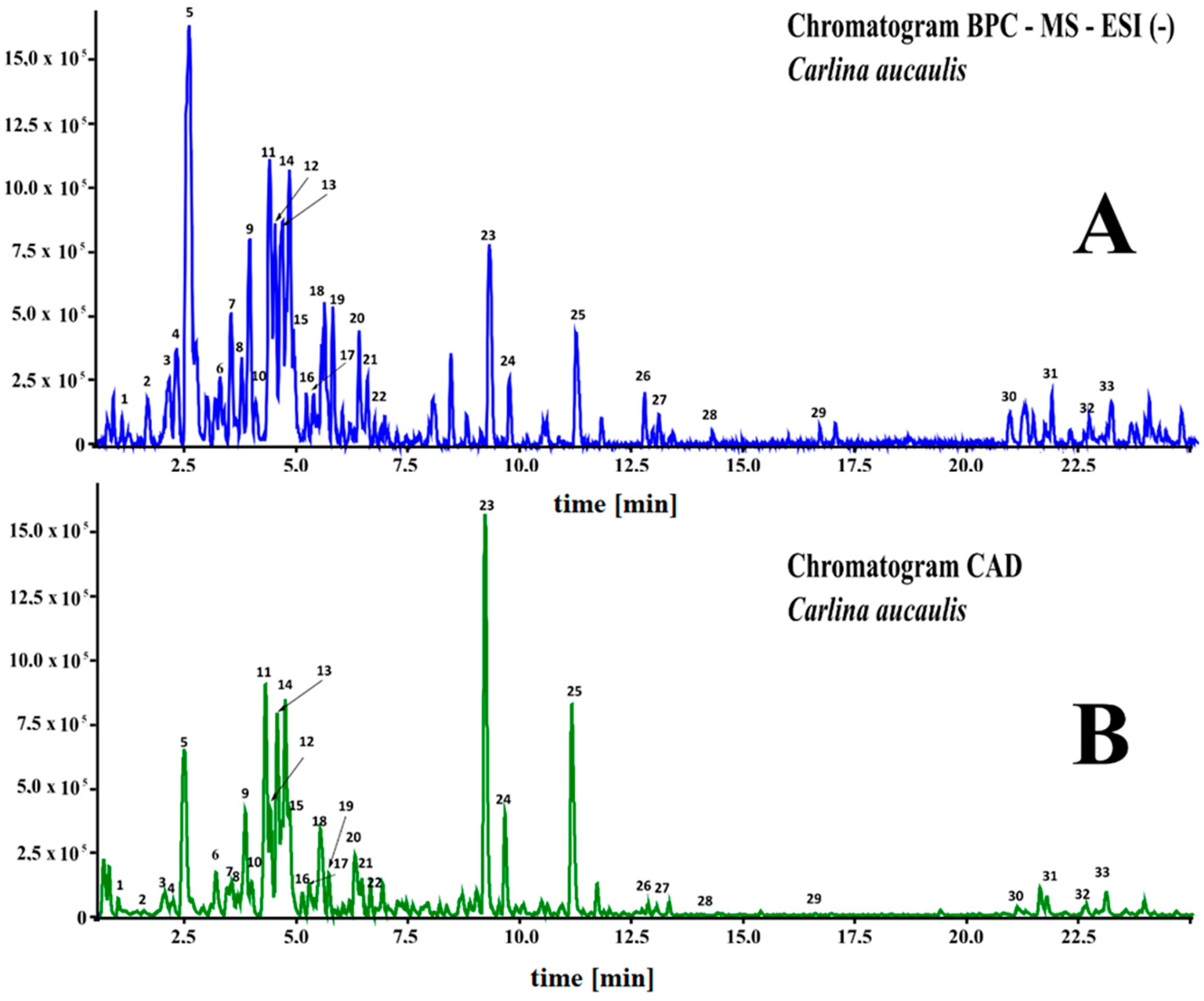
2.1. UHPLC-DAD-MS Analysis

Methanolic/water extracts from the above-ground parts of C. acaulis were separated, and their chemical composition was analyzed using UHPLC-MS. The compounds were identified based on mass data (m/z-H), fragmentation patterns, UV-Vis spectra, and additionally by comparison with standards when available. An example of the obtained chromatograms is shown in Figure 2.
A total of twenty different phenolic compounds were identified in the C. acaulis extract, including phenolic acids and flavonoids. Among the acids, 5-caffeoylquinic and 3-caffeoylquinic acids were the most abundant, with contents reaching up to 9.21 and 3.49 mg/g of dry weight, respectively. This was followed by dihydroxybenzoic acid (2.14 mg/g), quinic acid (0.99 mg/g), and 5-p-coumaroylquinic acid (0.57 mg/g).
From the flavonoid class, kaempferol dihexoside pentose (3.14 mg/g) was predominant, followed by C-glycosides of apigenin, namely shaftoside (6-C-glucoside-8-C-arabinoside), present at the highest amount, reaching 3.12 mg/g, isoschaftoside (6-C-arabinoside-8-C-glucoside) with a total content of 2.28 mg/g, vitexin-2-O-rhamnoside (1.96 mg/g), and isovitexin 2″-O-rhamnoside (1.74 mg/g). In addition, two derivatives of isovitexin, vicenin II and isovicenin, were found to have a content of 1.92 and 1.0 mg/g, respectively. The other flavonoids found in the aboveground part of C. acaulis were C-glycosides of luteolin, represented by isoorientin (6-C-glucoside), orientin (8-C-glucoside), 6-C-xylosyl luteolin, luteolin di-C-glucoside, and 2″-isoorientin O-glucopyranoside, with mean contents of 1.84, 1.05, 0.97, 0.69, and 0.41 mg/g, respectively. A small quantity of gossypetin dihexose (0.57 mg/g) and hesperitin 7-O-diglucosorhamnoside (0.2 mg/g) were also detected. Furthermore, several lipophilic constituents, including phospholipids 9,10-dihydroxy-8-oxooctadec-12-enoic acid, traumatic acid, and pinellic acid, are visible on the chromatogram of the methanol/water extract of C. acaulis.
The results of qualitative analysis and quantification, expressed per gram of dried plant material, are shown in Table 1.

2.2. Plant Material and Fractionation

Two years field cultivation of C. acaulis yielded 6.47 kg of plant material. After freeze-drying, the total weight of the lyophilized powder was 1.1 kg. Exhaustive extraction of 1 kg of freeze-dried samples with 70% methanol yielded 202 g of the first fraction (ECA).
ECA was subsequently fractionated using solvents with different polarities, including hexane, followed by ethyl acetate, and n-butanol, which yielded 5.62 g (HCA), 2.52 g (EaCA), and 5.26 g (BCA) of dried extract, respectively. An amount of 186 g of solid residues remaining after organic solvent extraction were dissolved in water (H2OCA).

2.2.1. Chromatographic Analysis

The qualitative and quantitative phytochemical composition of the extracts was characterized, and the results were expressed per g of dried extract (Table 2). Examples of the UHPLC-MS and PDA chromatograms obtained for specific fractions are shown in Figure S1 (Supplementary Material).
Chromatographic analysis revealed that the ethyl acetate fraction was predominant in phenolic compounds, which constitute approximately 55% of the dry weight of the extract. The butanol and water fractions contained significantly lower amounts of polyphenols, approximately 11% and 1.1% of the dry weight, respectively. Phospholipids remained in the hexane fraction.

2.2.2. Antioxidant Activity

The content of polyphenols in EaCA significantly affects free radical scavenging activity (DPPH) and ferric reducing power (FRAP) (Table 3). EaCA was the most potent free radical scavenger and had the highest ability to reduce ferric ions. BCA and H2OCA exhibited an almost equal effect, and HCV had the lowest antioxidant potential.

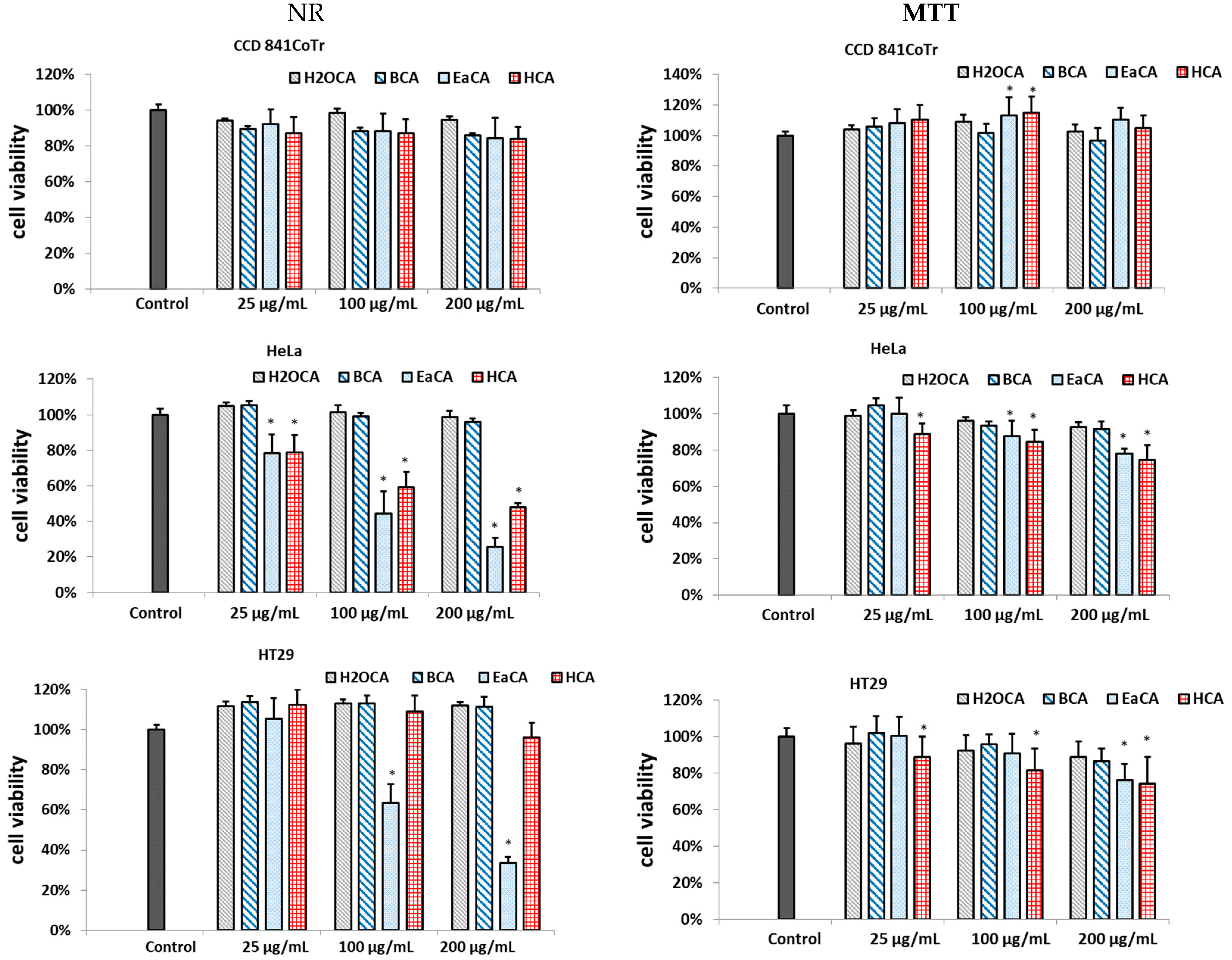
2.2.3. Cytotoxic Activity

In the study, two complementary tests were used to evaluate the cytotoxicity of the analyzed extracts against human colorectal adenocarcinoma (HT29) and human cervical cancer cells (HeLa), including MTT and the neutral red (NR) uptake assay. The MTT assesses cell metabolic activity based on the measurement of NAD(P)H-dependent cellular oxidoreductase. In turn, NR allows the detection of viable cells because the dye is retained in lysosomes in living cells. A normal epithelial colon cell line (CCD 841CoTr) was used as a reference line. The results are shown in Figure 3. The data indicated that all extracts at the tested concentration range were not toxic or showed mild toxicity against the reference line. In turn, EaCA and HCA exhibited cytotoxicity against HeLa, and EaCA also effectively reduced the number of viable cells in the HT29 line. For example, at a concentration of 200 µg/mL, it decreased the number of visible cells to 33% and 26% in HT29 and HeLa, respectively, while in CCD 841CoTr, viability stayed at the level of 84% (NR test). No effects were noted for BCa or H2OCA.

2.3. Bioactivity-Guided Subfractionation

2.3.1. Chromatographic Analysis

The EaCA fraction, being the most active against the tested cancer lines, was chosen for further fractionation using 20% methanol (EaCA_1), followed by 60% methanol (EaCA_2), 80% methanol (EaCA_3), and 100% methanol (EaCA_4). Phytochemical analysis (Table 4) revealed that phenolic acids were accumulated in the first fraction, constituting approximately 43% of the total yield. In contrast, the second fraction was abundant in flavonoid compounds (approximately 68% of the total yield). EaCA_3 contained fatty acids, including traumatic, pinellic, and 9,10-dihydroxy-8-oxsooctadec-12-enic acid (40.1%), and no polyphenolic constituents were found in the fraction. In EaCA_4, no components were identified because they were not detected by MS (they were not ionizable) or UV-Vis detectors (they had no chromophores).

2.3.2. Antioxidant Activity

As expected, EaCA_1 and EaCA_2 had the highest antioxidant activity because polyphenolic compounds were exhaustively extracted in the first two stages of the process. On the other hand, EaCA_3 and EaCA_4 had minimal free radical scavenging capacity and the ability to reduce ferric ions (Table 5).

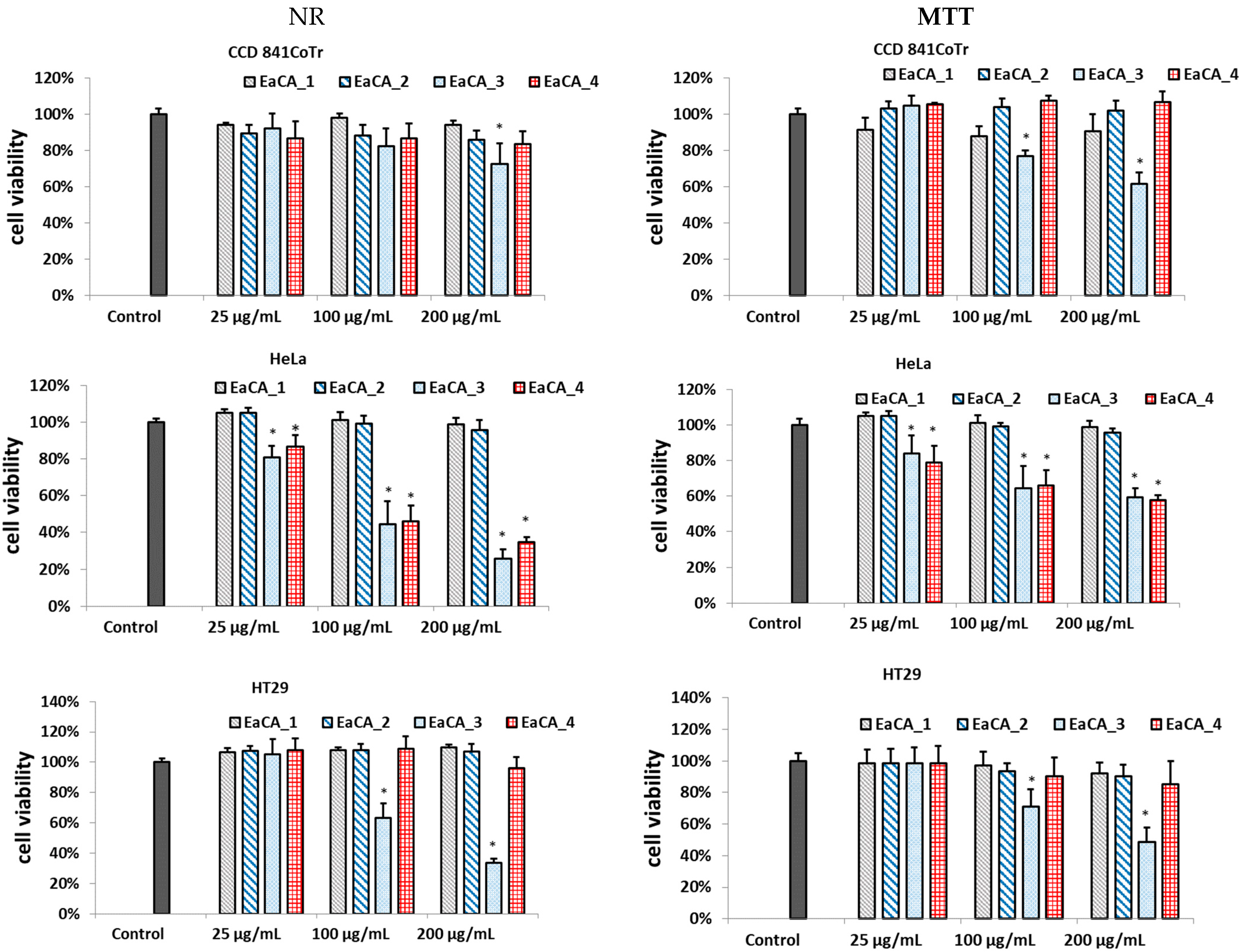
2.3.3. Cytotoxic Activity

The subfractions were analyzed in terms of cytotoxicity, and the results for the MTT and NR assays are shown in Figure 4.
As can be seen, the subfractions rich in polyphenolic compounds, EaCA_1 and EaCA_2, showed no cytotoxic effect in the tested cell lines. The most active subfraction was EaCA_3, in which fatty acids were detected. It showed the highest activity against HT29 and HeLa cancer lines; however, it also exhibits moderate cytotoxicity against normal epithelial colon cell lines. MTT assay showed that at a concentration of 100 µg/mL, it reduced cell viability to 64%, 71%, and 79% in HeLa, HT29, and CCD 841CoTr, respectively. In the NR test, the values were as follows: 44%, 63%, and 82% in HeLa, HT29, and CCD 841CoTr, respectively.
In turn, EaCA_4 did not negatively affect cell viability and metabolism in CCD 841CoTr. It effectively reduced both parameters in human cervical cancer cells; however, it had no impact on the HT29 line.

3. Discussion

Carlina acaulis is a plant with great significance in folk medicine; however, it is still poorly known in the context of phytochemical composition. UHPLC-DAD-MS analysis has revealed that the above-ground parts of C. acaulis are abundant in phenolic acids as well as flavonoids, mostly belonging to C-glycosides, which seem to be characteristic for the Carlina genus [20]. Some of the detected compounds, namely chlorogenic acid, schaftoside, homoorientin, orientin, vitexin, apigenin, and its 7-glucoside, were previously found in C. acaulis [13]; however, it should be noted that the identification was based on a comparison of retention time and UV-VIS spectrum with the standard. The present study not only confirmed these findings using mass data but also showed that C. acaulis contains a high amount of other polyphenolic compounds such as dihydroxybenzoic acid, vicenin II, vitexin, and orientin glycosides. Furthermore, it was shown that C. acaulis can serve as a rich source of C-glycosides, which are less widespread in nature than O-glycosides. These compounds possess the glycoside moiety attached directly to the flavonoid backbone through C-C covalent bonds, which makes their structure more stable and less susceptible to hydrolysis. This characteristic feature has an impact on their biological properties [21,22]. Some interesting effects have been found for these types of compounds. For example, schaftoside can inhibit the SARS-CoV-2 virus [23], vicenin-II was active against the Helicobacterium pylori infection [24] and may prevent osteoarthritis due to inhibition of extracellular matrix degradation [25], and orientin showed neuroprotective, vasodilatation, and cardioprotective activities [26].
Despite the wide application of C. acaulis in ethnomedicine, the number of scientific reports on the biological activity of this plant is scarce, and they are mainly focused on carlina oxide, the main volatile component of the essential oil. The present study revealed that C. acaulis extract showed cytotoxic potential against human colon cancer cells (HT29) and human cervical cancer cells (HeLa), with a mild cytotoxic effect on normal cell lines. In previous work, the cytotoxicity of this plant was also noted in human malignant melanoma [14].
It was expected that the activity was due to the high polyphenol content because many components identified in the extract showed anticancer potential in in vitro assays. For example, it has been evidenced that chlorogenic acids, which were abundant components of the extract, were effective against human oral squamous carcinoma (HSC-2) and salivary gland tumor (HSG) [27]. In addition, it reduced the proliferation rate, migration/invasion ability, and mitochondrial ATP production in human hepatoma and lung cancer cells [28]. In turn, vitexin and isovitexin decreased the viability of colorectal adenocarcinoma cells [29], and orientin was effective against human bladder carcinoma [30], breast cancer [31], and colorectal carcinoma [32]. Additionally, numerous other flavonoids have demonstrated potential as anticancer agents [33,34,35].
Surprisingly, the activity was not linked to the polyphenol content, as EaCA_1 and EaCA_2 subfractions, which were rich in these components, were not active. On the other hand, subfraction EaCA_3 was highly cytotoxic against both cancer lines and also showed toxicity against normal cells. This may indicate that the presence of polyphenols in the ethyl acetate fraction (EaCA) ameliorated the cytotoxicity of components from EaCA_3 because EaCA was not toxic against normal cells and retained activity toward cancer lines. Such protective activity was previously noted for chlorogenic acid against doxorubicin-induced cytotoxicity in cardiomyocytes [36].
Only three components were identified in the most cytotoxic EaCA_3, including traumatic acid (TA), 9,10-dihydroxy-8-oxooctadec-12-enoic acid, and pinellic acid (PA), which comprise approximately 40.1% of the total yield. Among them, TA may be responsible for the cytotoxic properties of the fraction, as this type of activity of TA was previously reported. TA is an oxidative derivative of unsaturated fatty acids synthesized from linoleic or linolenic acid during plant injury. It plays a role in the protection and repair of plant tissue. Its anticancer activity was described in the literature [37].
Jabłońska-Trypuć et al. have shown that TA is effective against breast cancer cells while simultaneously stimulating proliferation in healthy mammary epithelial cells [38]. It has also been shown that TA exhibits an antioxidant effect in normal human fibroblasts and stimulates ROS in breast cancer cells [39]. Increased ROS production in cells leads to oxidative stress, which, in turn, triggers lipid peroxidation—a detrimental process causing structural modifications to lipid-protein complexes [40]. Literature data have shown that cancer cells are more vulnerable to oxidation compared with normal cells [41,42]. Additionally, it has been found that TA disrupts the GSH/GSSG ratio, an essential antioxidant regulatory system, and induces apoptosis through the caspase pathway [43].
Moreover, there are also some research papers that have documented the cytotoxic properties of pinellic acid against cancer cell lines, specifically HepG2 and PC3 cell lines [44,45].
It should also be noted that the cytotoxic effect of the EaCA_3 subfraction may also be a result of the presence of other components that were not detected by MS and DAD. Therefore, further investigation is needed to clarify this issue. The present study indicates that the anticancer potential of C. acaulis is a promising direction for more detailed study. It also justified the traditional use of this plant.

4. Materials and Methods

4.1. Reagents and Standards
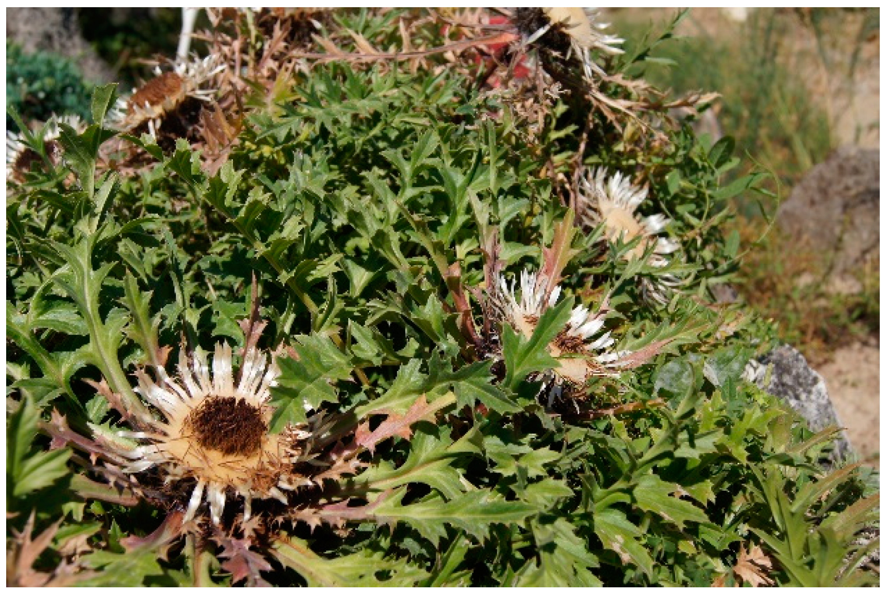
LC-MS-grade methanol, acetonitrile, and formic acid and reference standards were purchased from Merck (Sigma-Aldrich Co., St. Louis, MO, USA). The other solvents (Merck) were analytical grade. Water was purified using Ultrapure Milipore DirectQ 3UV-R (Merck KGaA, Darmstadt, Germany). Reagents, including 2.2-diphenyl-1-picrylhydrazyl (DPPH), o-phenanthroline, and ferric chloride (FRAP reagent), were purchased in Fluka (Sigma-Aldrich Co., St. Louis, MO, USA).

4.2. Plant Material

The field cultivation of C. acaulis was conducted following the protocol previously published by Strzemski et al. [46]. Seeds obtained from plants growing in the UMCS Botanical Garden (voucher specimen no. 2005A) were sown in early February in boxes filled with peat substrate. In mid-April, individual seedlings were transplanted into multipallets containing peat substrate. By the beginning of July, the plants had been transferred to loess soil at a density of four plants per square meter. Weeding was performed manually, and no fertilization or protective measures were applied during cultivation. Harvesting took place when the plants began flowering within the first ten days of July in the second year of cultivation.

4.3. Extraction and Fractionation

The above-ground parts of the plants were rinsed with running water, dried at room temperature for 48 h, and powdered using a laboratory grinder, IKA A11 (IKA-Werke, Stufen, Germany). Furthermore, the plant material was freeze-dried (0.001 mbar) for 72 h using a Christ Alpha 2-4 LDplus laboratory freeze dryer (Martin Christ Gefriertrocknungsanlagen GmbH, Osterode am Harz, Germany). An amount of 1000 g of dried material was exhaustively extracted with methanol and 70% methanol (3 × 5 L and 3 × 1.5 L for 15 min each) using an ultrasonic bath. The extracts (ECA) were combined, centrifuged at 8000 RPM, filtered, and then concentrated using a vacuum evaporator. They were then frozen and subjected to freeze-drying. The freeze-dried ECA was dissolved in MeOH and subjected to liquid-liquid extractions with n-hexane (5 × 100 mL), followed by ethyl acetate (5 × 100 mL). The resulting residue was evaporated to dryness, suspended in water, and then extracted with n-butanol (5 × 100 mL). The residue obtained after extraction represented the H2OCV fraction. The fractions were concentrated using a vacuum evaporator, frozen, and freeze-dried [20]. Based on the bioactivity assay, the most active fraction (ethyl acetate) was subjected to further fractionation. The ethyl acetate fractions were dissolved in deionized water with the addition of 5% DMSO and then centrifuged (1000× g; 10 min). The supernatant was loaded onto a glass preparative column with dimensions of 10 × 30 cm, filled with Cosmosil 75C18—PREP chromatographic packing material (75 μm, Nacalai Tesque), previously impregnated with 1% MeOH in water. Approximately 10 g of the dissolved extract was loaded onto the column at once, and elution was performed using a stepwise gradient of the mobile phase with 3 times the column volume for each concentration. The elution used appropriate amounts of 20%, 60%, and 100% MeOH to obtain the following fractions: EaCA_1 (20% MeOH), EaCA_2 (60% MeOH), EaCA_3 (80% MeOH), and EaCA_4 (100% MeOH).

4.4. Chromatographic Analysis (UHPLC—HR/QTOF/MS—CAD–PDA)

The lyophilized samples were dissolved in a mixture of 50% MeOH in water with the addition of 5% dimethyl sulfoxide (DMSO) and then centrifuged and filtered. Chromatographic separation and qualitative analyses were performed using an ultra-high-performance liquid chromatograph (UHPLC) coupled with a charged aerosol detector (CAD) and a high-resolution/quadropole time-of-flight mass spectrometer (HR/QTOF/MS Impact II) equipped with an electrospray ionization (ESI) source. The analyses were carried out on a BEH C18 column (2.1 × 150 mm, 1.7 µm; Waters) at a temperature of 40 °C. The mobile phase consisted of a mixture of acetonitrile with 0.1% formic acid (FA) (solvent B) and water with 0.1% formic acid (FA) (solvent A). A linear gradient elution was performed as follows: from 2% B to 80% B over a period of 30 min, at a flow rate of 0.5 mL/min. MS spectra were acquired in the range of 80–2000 m/z in negative ionization. The working MS parameters were as previously described [20]. The Waters Mass Lynx software was used to process the data. Quantification was carried out using an ultra-performance liquid chromatograph combined with a photodiode detector and a triple quadrupole mass spectrometer (UPLC-PDA-ESI-MS) (ACQUITY TQD, Waters). UV-Vis spectra of the components were registered in the range of 190–750 nm (resolution of 3.6 nm).

4.5. Cell Cultures

The HT29 cell line (ATCC® No. HTB-38™) is a human colorectal adenocarcinoma cell line (grade I), derived from a 44-year-old female adult. These cells were cultured in RPMI 1640 medium supplemented with 10% fetal calf serum (FCS) (GibcoTM, Paisley, UK) and antibiotics (100 U/mL penicillin, 100 μg/mL streptomycin, and 0.25 μg/mL amphotericin B) (GibcoTM, Paisley, UK) at 37 °C in a humidified atmosphere with 5% CO2.
The CCD 841 CoTr cell line (ATCC® No. CRL-1807™) is a human normal colon epithelial cell line (SV40 transformed), derived from a female at 21 weeks of gestation. These cells were cultured in RPMI 1640 + DMEM (1:1) medium (Sigma-Aldrich Co. LLC, St. Louis, MO, USA) supplemented with 10% FCS and antibiotics at 37 °C in a humidified atmosphere with 5% CO2. The human cervical carcinoma cell line (HeLa, ECACC No. 85060701) was also used in this study. Cells were grown as monolayers in 25-cm3 culture flasks (Nunc., Roskilde, Denmark) in RPMI 1640 medium supplemented with 5% fetal bovine serum (FBS) (Gibco, Paisley, UK) and antibiotics (100 U/mL penicillin, 100 µg/mL streptomycin) (Gibco, Paisley, UK) at 37 °C in a humidified atmosphere with 5% CO2. For cytotoxicity assay, the cells (1 × 10⁵ cells/mL) were plated in 96-well flat-bottom plates, incubated for 24 h at 37 °C, and then treated with the fractions and subfractions for 24 h. Stock solution was prepared using DMSO/culture medium (1:1) and appropriately diluted. The final concentration of DMSO did not exceed 0.5%. Cells treated with 0.5% DMSO in culture medium were taken as controls.
All experiments were performed in triplicate for each extract concentration and presented as a percentage of the control (100%).

4.6. Cell Viability Assay

4.6.1. MTT Assay

After incubating the samples for 24 h, a solution of 3-(4,5-dimethylthiazole-2-yl)-2,5-diphenyltetrazolium bromide (MTT) at a concentration of 5 mg/mL (Sigma) was introduced to the cells (25 μL/well), followed by an additional 3-h incubation. The resulting insoluble formazan crystals were dissolved overnight in a mixture containing 10% sodium dodecyl sulfate (SDS) in 0.01 M HCl. The absorbance was gauged at a wavelength of 570 nm using an E-max Microplate Reader (Molecular Devices Corporation, Menlo Park, CA, USA) [47].

4.6.2. Neutral Red Uptake Assay

The procedure was based on Zarandi et al. with minor modifications [48]. Following a 24-h incubation period with the samples, the liquid in the wells was discarded, and the cells were subjected to a 2-h incubation with a neutral red dye solution (40 μg/mL) at 37 °C. Subsequently, the cells were rinsed with phosphate-buffered saline (PBS), and after removing the PBS, decolorizing buffer (150 µL) was introduced. The plates were agitated for 10 min, and the optical density (OD) of the extracted dye was assessed at 540 nm using an E-max Micro-plate Reader (Molecular Devices Corporation, Menlo Park, CA, USA). The outcomes are depicted as the percentage of retained dye quantity in comparison to the control cells (100%).

4.7. Antioxidant Activity

4.7.1. DPPH• Free Radical Scavenging Test

The free radical scavenging activity of the tested compounds was evaluated using the 1,1-diphenyl-2-picrylhydrazyl (DPPH•) assay [49]. This method is based on the antioxidant’s ability of to reduce the stable dark violet DPPH• radical (Sigma) to the yellow diphenyl-picrylhydrazine. Briefly, 100 μL of DPPH• solution (0.2 mg/mL in ethanol) was added to 100 μL of various concentrations of the tested compounds (ranging from 0 to 200 μg/mL) as well as to standard Trolox (Sigma) at increasing concentrations (1–50 μg/mL) used as a reference for free radical scavenging activity. After incubating the samples for 20 min at room temperature, the absorbance of the solutions was measured at 515 nm using a microplate reader (BioTek Instruments, Winooski, VT, USA). A lower absorbance value indicates a higher free radical scavenging activity of the compounds. The activity of each substance was determined by comparing its absorbance with that of a blank solution (containing reagents without the compound) and the standard. The capability of each substance to scavenge the DPPH• radical was then calculated using the following formula:
DPPH• scavenging effect (%) = [(X control − X substance/X control) × 100]
where X control is the absorbance of the control and X substance is the absorbance in the presence of tested compound.

4.7.2. Ferric-Reducing Antioxidant Power Assay (FRAP)

Each tested compound concentration was dissolved in Milli-Q water and then mixed with an equal volume of 0.2 M sodium phosphate buffer (pH 6.6) and 1% potassium ferricyanide. The mixture was incubated for 30 min at 37 °C. After the incubation, 10% trichloroacetic acid (w/v) was added to the mixture, and the resulting solution was centrifuged at 1000× g for 5 min. Next, 1 mL of the upper layer was mixed with an equal volume of Milli-Q water and 0.1% ferric chloride. The absorbance of the solution was measured at 700 nm using a spectrophotometer (BioTek Instruments, Winooski, VT, USA). Ascorbic acid was used as the positive control in this assay [20].

4.8. Statistical Analysis

All analyses were conducted three times. The obtained data were subjected to statistical analysis using Statistic version 13.3 software. One-way ANOVA was performed, followed by Dunnett’s post hoc test. The results were presented as the mean ± standard deviation (SD). Statistical significance was determined at a p-value of ≤ 0.05.

5. Conclusions

In the present work, the phytochemical composition and cytotoxic potential of C. acaulis extract against human colorectal adenocarcinoma (HT29) and human cervical cancer (HeLa) cells were investigated. The investigation revealed that C. acaulis is a rich source of phenolic acids and flavonoids, with predominant chlorogenic acids and C-glycosides of luteolin and apigenin. Among the flavonoids, kaempferol dihexosidipentose and schaftoside were the most abundant, followed by isoorientin, vitexin-2-O-rhamnoside, and vicenin II. Furthermore, it has been demonstrated that C. acaulis shows significant cytotoxicity against the tested cell lines. However, the effect was not linked to the presence of polyphenolic compounds. The subfraction containing oxylipins belonging to oxygenated fatty acids was the most active and decreased the viability of HeLa and HT29 cells with minimal negative effects on normal cells.
The present study indicates that the anticancer potential of C. acaulis is a promising direction for more detailed study. In the future, the work should be focused on exploring the molecular mechanisms connected with the biological activity of the plant. Furthermore, the isolation of specific components should be carried out to identify the component with a specific biological effect.

Supplementary Materials

The following supporting information can be downloaded at: https://www.mdpi.com/article/10.3390/molecules28166148/s1. Figure S1: Chromatograms obtained from ultra-performance liquid chromatography with mass spectrometry and electrospray ionization UHPLC-ESI-MS(-) (a) and UHPLC-ESI-MS (+) (b) and UHPLC with photodiode detector—PDA (254 nm) (c). These chromatograms represent fractions obtained through liquid–liquid extraction from the extract of C. acaulis.

Author Contributions

Conceptualization. M.W., J.M. and I.S.; methodology. M.W., J.M., R.P. and I.S.; validation. M.W., J.M., R.P. and I.S.; formal analysis. M.W., R.P., J.M. and I.S; investigation. M.W., J.M., R.P., M.S., K.T.-R., D.S., S.D., J.S. and I.S.; writing—original draft preparation. M.W. and I.S.; writing—review and editing. M.W. and I.S.; visualization. S.D., J.M. and M.S.; supervision. M.W. and I.S. All authors have read and agreed to the published version of the manuscript.

Funding

This research received no external funding.

Institutional Review Board Statement

Not applicable.

Informed Consent Statement

Not applicable.

Data Availability Statement

Data are contained within the article or Supplementary Materials.

Conflicts of Interest

The authors declare no conflict of interest.

References

  1. Spinozzi, E.; Ferrati, M.; Cappellacci, L.; Caselli, A.; Perinelli, D.R.; Bonacucina, G.; Maggi, F.; Strzemski, M.; Petrelli, R.; Pavela, R.; et al. Carlina acaulis L. (Asteraceae): Biology, Phytochemistry, and Application as a Promising Source of Effective Green Insecticides and Acaricides. Ind. Crops Prod. 2023, 192, 116076. [Google Scholar] [CrossRef]
  2. Strzemski, M.; Wójciak-Kosior, M.; Sowa, I.; Załuski, D.; Verpoorte, R. Historical and Traditional Medical Applications of Carlina acaulis L.—A Critical Ethnopharmacological Review. J. Ethnopharmacol. 2019, 239, 111842. [Google Scholar] [CrossRef] [PubMed]
  3. Rexhepi, B.; Mustafa, B.; Hajdari, A.; Rushidi-Rexhepi, J.; Quave, C.L.; Pieroni, A. Traditional Medicinal Plant Knowledge among Albanians, Macedonians and Gorani in the Sharr Mountains (Republic of Macedonia). Genet. Resour. Crop Evol. 2013, 60, 2055–2080. [Google Scholar] [CrossRef]
  4. Guarrera, P.M. Food Medicine and Minor Nourishment in the Folk Traditions of Central Italy (Marche, Abruzzo and Latium). Fitoterapia 2003, 74, 515–544. [Google Scholar] [CrossRef]
  5. Jarić, S.; Popović, Z.; Mačukanović-Jocić, M.; Djurdjević, L.; Mijatović, M.; Karadžić, B.; Mitrović, M.; Pavlović, P. An Ethnobotanical Study on the Usage of Wild Medicinal Herbs from Kopaonik Mountain (Central Serbia). J. Ethnopharmacol. 2007, 111, 160–175. [Google Scholar] [CrossRef] [PubMed]
  6. Šarić-Kundalić, B.; Dobeš, C.; Klatte-Asselmeyer, V.; Saukel, J. Ethnobotanical Study on Medicinal Use of Wild and Cultivated Plants in Middle, South and West Bosnia and Herzegovina. J. Ethnopharmacol. 2010, 131, 33–55. [Google Scholar] [CrossRef]
  7. Wnorowska, S.; Targowska-Duda, K.; Kurzepa, J.; Wnorowski, A.; Strzemski, M. Carlina Oxide Inhibits the Interaction of SARS-CoV-2 S Glycoprotein with Angiotensin-Converting Enzyme 2. Ind. Crops Prod. 2022, 187, 115338. [Google Scholar] [CrossRef] [PubMed]
  8. Wnorowski, A.; Wnorowska, S.; Wojas-Krawczyk, K.; Grenda, A.; Staniak, M.; Michalak, A.; Woźniak, S.; Matosiuk, D.; Biała, G.; Wójciak, M.; et al. Toxicity of Carlina Oxide—A Natural Polyacetylene from the Carlina acaulis Roots—In Vitro and in Vivo Study. Toxins 2020, 12, 239. [Google Scholar] [CrossRef]
  9. Kavallieratos, N.G.; Nika, E.P.; Skourti, A.; Spinozzi, E.; Ferrati, M.; Petrelli, R.; Maggi, F.; Benelli, G. Carlina acaulis Essential Oil: A Candidate Product for Agrochemical Industry Due to Its Pesticidal Capacity. Ind. Crops Prod. 2022, 188, 115572. [Google Scholar] [CrossRef]
  10. Herrmann, F.; Hamoud, R.; Sporer, F.; Tahrani, A.; Wink, M. Carlina Oxide—A Natural Polyacetylene from Carlina acaulis (Asteraceae) with Potent Antitrypanosomal and Antimicrobial Properties. Planta Med. 2011, 77, 1905–1911. [Google Scholar] [CrossRef]
  11. Benelli, G.; Pavoni, L.; Zeni, V.; Ricciardi, R.; Cosci, F.; Cacopardo, G.; Gendusa, S.; Spinozzi, E.; Petrelli, R.; Cappellacci, L.; et al. Developing a Highly Stable Carlina acaulis Essential Oil Nanoemulsion for Managing Lobesia Botrana. Nanomaterials 2020, 10, 1867. [Google Scholar] [CrossRef] [PubMed]
  12. Benelli, G.; Pavela, R.; Petrelli, R.; Nzekoue, F.K.; Cappellacci, L.; Lupidi, G.; Quassinti, L.; Bramucci, M.; Sut, S.; Dall’Acqua, S.; et al. Carlina Oxide from Carlina acaulis Root Essential Oil Acts as a Potent Mosquito Larvicide. Ind. Crops Prod. 2019, 137, 356–366. [Google Scholar] [CrossRef]
  13. Dordević, S.; Tadić, V.; Petrović, S.; Kukić-Marković, J.; Dobrić, S.; Milenković, M.; Hadžifejzović, N. Bioactivity Assays on Carlina acaulis and C. Acanthifolia Root and Herb Extracts. Dig. J. Nanomater. Biostruct. 2012, 7, 1213–1222. [Google Scholar]
  14. Strzemski, M.; Wojnicki, K.; Sowa, I.; Wojas-Krawczyk, K.; Krawczyk, P.; Kocjan, R.; Such, J.; Latalski, M.; Wnorowski, A.; Wójciak-Kosior, M. In Vitro Antiproliferative Activity of Extracts of Carlina acaulis Subsp. Caulescens and Carlina Acanthifolia Subsp. Utzka. Front. Pharmacol. 2017, 8, 371. [Google Scholar] [CrossRef]
  15. Stojanović-Radić, Z.; Čomić, L.; Radulović, N.; Blagojević, P.; Mihajilov-Krstev, T.; Rajković, J. Commercial Carlinae radix Herbal Drug: Botanical Identity, Chemical Composition and Antimicrobial Properties. Pharm. Biol. 2012, 50, 933–940. [Google Scholar] [CrossRef]
  16. Kavallieratos, N.G.; Nika, E.P.; Skourti, A.; Boukouvala, M.C.; Ntalaka, C.T.; Maggi, F.; Spinozzi, E.; Petrelli, R.; Perinelli, D.R.; Benelli, G.; et al. Carlina acaulis Essential Oil Nanoemulsion as a New Grain Protectant against Different Developmental Stages of Three Stored-product Beetles. Pest Manag. Sci. 2022, 78, 2434–2442. [Google Scholar] [CrossRef]
  17. Strzemski, M.; Wójciak-Kosior, M.; Sowa, I.; Rutkowska, E.; Szwerc, W.; Kocjan, R.; Latalski, M. Carlina Species as a New Source of Bioactive Pentacyclic Triterpenes. Ind. Crops Prod. 2016, 94, 498–504. [Google Scholar] [CrossRef]
  18. Jaiswal, R.; Deshpande, S.; Kuhnert, N. Profiling the chlorogenic acids of Rudbeckia hirta, Helianthus tuberosus, Carlina acaulis and Symphyotrichum novae-angliae leaves by LC-MSn. Phytochem. Anal. 2011, 22, 432–441. [Google Scholar] [CrossRef]
  19. Raynaud, J.; Rasolojaona, L. Flavonoïdes Des Feuilles de Carlina acaulis. Planta Med. 1979, 37, 168–171. [Google Scholar] [CrossRef]
  20. Sowa, I.; Mołdoch, J.; Dresler, S.; Kubrak, T.; Soluch, A.; Szczepanek, D.; Strzemski, M.; Paduch, R.; Wójciak, M. Phytochemical Profiling, Antioxidant Activity, and Protective Effect against H2O2-Induced Oxidative Stress of Carlina Vulgaris Extract. Molecules 2023, 28, 5422. [Google Scholar] [CrossRef]
  21. Xie, L.; Deng, Z.; Zhang, J.; Dong, H.; Wang, W.; Xing, B.; Liu, X. Comparison of Flavonoid O-Glycoside, C-Glycoside and Their Aglycones on Antioxidant Capacity and Metabolism during In Vitro Digestion and In Vivo. Foods 2022, 11, 882. [Google Scholar] [CrossRef] [PubMed]
  22. Xiao, J.; Capanoglu, E.; Jassbi, A.R.; Miron, A. Advance on the Flavonoid C-Glycosides and Health Benefits. Crit. Rev. Food Sci. Nutr. 2016, 56 (Suppl. 1), S29–S45. [Google Scholar] [CrossRef] [PubMed]
  23. Yi, Y.; Zhang, M.; Xue, H.; Yu, R.; Bao, Y.-O.; Kuang, Y.; Chai, Y.; Ma, W.; Wang, J.; Shi, X.; et al. Schaftoside Inhibits 3CLpro and PLpro of SARS-CoV-2 Virus and Regulates Immune Response and Inflammation of Host Cells for the Treatment of COVID-19. Acta Pharm. Sin. B 2022, 12, 4154–4164. [Google Scholar] [CrossRef]
  24. Zhang, Y.; Sun, J.; Dong, Y.; Shen, X.; Zhang, Z. Vicenin-2 Inhibits the Helicobacterium pylori Infection Associated Gastric Carcinogenic Events through Modulation of PI3K/AKT and Nrf2 Signaling in GES-1 Cells. J. Biochem. Mol. Toxicol. 2021, 35. [Google Scholar] [CrossRef]
  25. Chen, Y.-Y.; Yan, X.-J.; Jiang, X.-H.; Lu, F.-L.; Yang, X.-R.; Li, D.-P. Vicenin 3 Ameliorates ECM Degradation by Regulating the MAPK Pathway in SW1353 Chondrocytes. Exp. Ther. Med. 2021, 22, 1461. [Google Scholar] [CrossRef] [PubMed]
  26. Lam, K.Y.; Ling, A.P.K.; Koh, R.Y.; Wong, Y.P.; Say, Y.H. A Review on Medicinal Properties of Orientin. Adv. Pharmacol. Sci. 2016, 2016, 1–9. [Google Scholar] [CrossRef]
  27. Jiang, Y.; Kusama, K.; Satoh, K.; Takayama, F.; Watanabe, S.; Sakagami, H. Induction of Cytotoxicity by Chlorogenic Acid in Human Oral Tumor Cell Lines. Phytomedicine 2000, 7, 483–491. [Google Scholar] [CrossRef]
  28. Huang, S.; Wang, L.-L.; Xue, N.-N.; Li, C.; Guo, H.-H.; Ren, T.-K.; Zhan, Y.; Li, W.-B.; Zhang, J.; Chen, X.-G.; et al. Chlorogenic Acid Effectively Treats Cancers through Induction of Cancer Cell Differentiation. Theranostics 2019, 9, 6745–6763. [Google Scholar] [CrossRef]
  29. Tremmel, M.; Kiermaier, J.; Heilmann, J. In Vitro Metabolism of Six C-Glycosidic Flavonoids from Passiflora incarnata L. IJMS 2021, 22, 6566. [Google Scholar] [CrossRef]
  30. Tian, F.; Tong, M.; Li, Z.; Huang, W.; Jin, Y.; Cao, Q.; Zhou, X.; Tong, G. The Effects of Orientin on Proliferation and Apoptosis of T24 Human Bladder Carcinoma Cells Occurs Through the Inhibition of Nuclear Factor-KappaB and the Hedgehog Signaling Pathway. Med. Sci. Monit. 2019, 25, 9547–9554. [Google Scholar] [CrossRef]
  31. Kim, S.-J.; Pham, T.-H.; Bak, Y.; Ryu, H.-W.; Oh, S.-R.; Yoon, D.-Y. Orientin Inhibits Invasion by Suppressing MMP-9 and IL-8 Expression via the PKCα/ERK/AP-1/STAT3-Mediated Signaling Pathways in TPA-Treated MCF-7 Breast Cancer Cells. Phytomedicine 2018, 50, 35–42. [Google Scholar] [CrossRef] [PubMed]
  32. Thangaraj, K.; Balasubramanian, B.; Park, S.; Natesan, K.; Liu, W.; Manju, V. Orientin Induces G0/G1 Cell Cycle Arrest and Mitochondria Mediated Intrinsic Apoptosis in Human Colorectal Carcinoma HT29 Cells. Biomolecules 2019, 9, 418. [Google Scholar] [CrossRef] [PubMed]
  33. Kopustinskiene, D.M.; Jakstas, V.; Savickas, A.; Bernatoniene, J. Flavonoids as Anticancer Agents. Nutrients 2020, 12, 457. [Google Scholar] [CrossRef] [PubMed]
  34. Ponte, L.G.S.; Pavan, I.C.B.; Mancini, M.C.S.; Da Silva, L.G.S.; Morelli, A.P.; Severino, M.B.; Bezerra, R.M.N.; Simabuco, F.M. The Hallmarks of Flavonoids in Cancer. Molecules 2021, 26, 2029. [Google Scholar] [CrossRef]
  35. Forni, C.; Rossi, M.; Borromeo, I.; Feriotto, G.; Platamone, G.; Tabolacci, C.; Mischiati, C.; Beninati, S. Flavonoids: A Myth or a Reality for Cancer Therapy? Molecules 2021, 26, 3583. [Google Scholar] [CrossRef]
  36. Salzillo, A.; Ragone, A.; Spina, A.; Naviglio, S.; Sapio, L. Chlorogenic Acid Enhances Doxorubicin-Mediated Cytotoxic Effect in Osteosarcoma Cells. IJMS 2021, 22, 8586. [Google Scholar] [CrossRef]
  37. Pietryczuk, A.; Biziewska, I.; Imierska, M.; Czerpak, R. Influence of Traumatic Acid on Growth and Metabolism of Chlorella Vulgaris under Conditions of Salt Stress. Plant Growth Regul. 2014, 73, 103–110. [Google Scholar] [CrossRef]
  38. Jabłońska-Trypuć, A.; Wydro, U.; Wołejko, E.; Butarewicz, A. Toxicological Effects of Traumatic Acid and Selected Herbicides on Human Breast Cancer Cells: In Vitro Cytotoxicity Assessment of Analyzed Compounds. Molecules 2019, 24, 1710. [Google Scholar] [CrossRef]
  39. Jabłońska-Trypuć, A.; Pankiewicz, W.; Czerpak, R. Traumatic Acid Reduces Oxidative Stress and Enhances Collagen Biosynthesis in Cultured Human Skin Fibroblasts. Lipids 2016, 51, 1021–1035. [Google Scholar] [CrossRef]
  40. Dias, I.H.K.; Milic, I.; Heiss, C.; Ademowo, O.S.; Polidori, M.C.; Devitt, A.; Griffiths, H.R. Inflammation, Lipid (Per)Oxidation, and Redox Regulation. Antioxid. Redox Signal. 2020, 33, 166–190. [Google Scholar] [CrossRef]
  41. Van Der Paal, J.; Neyts, E.C.; Verlackt, C.C.W.; Bogaerts, A. Effect of Lipid Peroxidation on Membrane Permeability of Cancer and Normal Cells Subjected to Oxidative Stress. Chem. Sci. 2016, 7, 489–498. [Google Scholar] [CrossRef] [PubMed]
  42. Clemente, S.M.; Martínez-Costa, O.H.; Monsalve, M.; Samhan-Arias, A.K. Targeting Lipid Peroxidation for Cancer Treatment. Molecules 2020, 25, 5144. [Google Scholar] [CrossRef] [PubMed]
  43. Jabłońska-Trypuć, A.; Krętowski, R.; Wołejko, E.; Wydro, U.; Butarewicz, A. Traumatic Acid Toxicity Mechanisms in Human Breast Cancer MCF-7 Cells. Regul. Toxicol. Pharmacol. 2019, 106, 137–146. [Google Scholar] [CrossRef] [PubMed]
  44. Samra, R.M.; Soliman, A.F.; Zaki, A.A.; Ashour, A.; Al-Karmalawy, A.A.; Hassan, M.A.; Zaghloul, A.M. Bioassay-Guided Isolation of a New Cytotoxic Ceramide from Cyperus rotundus L. S. Afr. J. Bot. 2021, 139, 210–216. [Google Scholar] [CrossRef]
  45. Siegel, I.; Liu, T.L.; Yaghoubzadeh, E.; Keskey, T.S.; Gleicher, N. Cytotoxic Effects of Free Fatty Acids on Ascites Tumor Cells. J. Natl. Cancer Inst. 1987, 78, 271–277. [Google Scholar]
  46. Strzemski, M.; Dzida, K.; Dresler, S.; Sowa, I.; Kurzepa, J.; Szymczak, G.; Wójciak, M. Nitrogen Fertilisation Decreases the Yield of Bioactive Compounds in Carlina acaulis L. Grown in the Field. Ind. Crops Prod. 2021, 170, 113698. [Google Scholar] [CrossRef]
  47. Ghasemi, M.; Turnbull, T.; Sebastian, S.; Kempson, I. The MTT Assay: Utility, Limitations, Pitfalls, and Interpretation in Bulk and Single-Cell Analysis. IJMS 2021, 22, 12827. [Google Scholar] [CrossRef]
  48. Kheirandish Zarandi, P.; Zare Mirakabadi, A.; Sotoodehnejadnematalahi, F. Cytotoxic and Anticancer Effects of ICD-85 (Venom Derived Peptides) in Human Breast Adenocarcinoma and Normal Human Dermal Fibroblasts. Iran. J. Pharm. Res. 2019, 18, 232–240. [Google Scholar]
  49. Baliyan, S.; Mukherjee, R.; Priyadarshini, A.; Vibhuti, A.; Gupta, A.; Pandey, R.P.; Chang, C.-M. Determination of Antioxidants by DPPH Radical Scavenging Activity and Quantitative Phytochemical Analysis of Ficus Religiosa. Molecules 2022, 27, 1326. [Google Scholar] [CrossRef]
Figure 1. An example of C. aculis growing in its natural habitat.
Molecules 28 06148 g001
Figure 2. Chromatograms of extracts from C. acaulis obtained using a quadrupole-time-of-flight high-resolution mass spectrometer (LC-HRMS-QTOF) and a charged aerosol detector (CAD). (A) MS chromatogram (electrospray ionization—ESI); (B) CAD chromatogram. Numbering of the compounds as shown in Table 1.
Molecules 28 06148 g002
Figure 3. Effect of the different concentrations of fractions obtained from methanol/water extract of Carlina acaulis on cell viability as determined by the NR and MTT assays and expressed as a % of control (0.5% of DMSO in medium). The fractions were obtained using hexane (HCA), followed by ethyl acetate (EaCA), butanol (BCA), and water (H2OCA). The data are means (n = 3) ± SD. One-way ANOVA followed by Dunnett’s post hoc test; the differences were considered significant at p < 0.05. * indicates statistically significant difference.
Molecules 28 06148 g003
Figure 4. Effect of the different concentrations of subfractions obtained from ethyl acetate extract of Carlina acaulis (EaCA) on cell viability determined by the NR and MTT assays expressed as a % of control (0.5% of DMSO in medium). The fractions were obtained using 20% methanol (EaCA_1), followed by 60% methanol (EaCA_2), 80% methanol (EaCA_3), and 100% methanol (EaCA_4). The data are means (n = 3) ± SD. One-way ANOVA followed by Dunnett’s post hoc test; the differences were considered significant at p < 0.05. * indicates statistically significant difference.
Molecules 28 06148 g004
Table 1. Compounds found in the extract of aerial parts of C. acaulis. The quantification is expressed per gram of dried material.
NrRT (min)M/ZMS2Ion Formula [M/Z-H]ΔppmIdentifiedAmount (mg/g DW)
10.8191.055878191, 135C7H11O61.2quinic acid0.99 ± 0.01
21.7153.019025153, 109C7H5O42.0dihydroxybenzoic acid2.14 ± 0.01
32.3343.102756343, 135C15H19O92.0unknown
42.5353.087070353, 351, 191, 133C16H17O92.13-caffeoylquinic acid3.49 ± 0.03
52.6353.087049353, 191C16H17O92.15-caffeoylquinic acid9.21 ± 0.01
63.0373.113574373, 165, 150C16H21O101.2unknown
73.3609.145133609, 489, 399, 369C27H29O161.6luteolin di-C-glucoside0.69 ± 0.01
83.5337.092161337, 191C16H17O82.25-p-coumaroylquinic acid0.57 ± 0.06
93.9593.150084593, 473, 383, 353C27H29O151.9vicenin II1.92 ± 0.01
104.3609.145243609, 489, 429, 357C27H29O161.42”-isoorientin O-glucopyranoside0.41 ± 0.01
114.4563.139696563, 473, 443, 383, 353C26H27O141.7shaftoside3.12 ± 0.01
124.6447.092277447, 429, 357, 327, 297C21H19O112.3orientin1.05 ± 0.01
134.8563.139620563, 503, 473, 443, 383, 353C26H27O141.8isoshaftoside (I)1.06 ± 0.01
144.9579.134349579, 459, 429, 357, 327, 309C26H27O152.1isoorientin1.84 ± 0.01
155.2563.139889563, 473, 443, 383, 353C26H27O141.3isoshaftoside (II)1.22 ± 0.01
165.3593.150261593, 473, 383, 293C27H29O151.6isovicenin1.00 ± 0.01
175.5417.081764417, 357, 327, 297C20H17O102.36-C-xylosyl luteolin0.97 ± 0.01
185.6577.155385577, 457, 413, 341, 311, 293C27H29O141.5vitexin-2-O-rhamnoside1.96 ± 0.01
195.8525.233345525, 481, 319, 301, 119C25H34O121.5lucidumoside A
206.3577.155674577, 457, 445, 427, 324C27H29O141.0isovitexin 2″-O-rhamnoside1.74 ± 0.01
216.4435.412847435, 273, 167C21H24O101.7phlorizin
226.5413.413442413, 269, 161C19H25O102.1unknown
239.2771.343701771,609, 489, 447, 343, 301C37H55O171.0hesperitin 7-O diglucosorhamnoside0.20 ± 0.01
249.7741.168209741, 285C32H38O201.3kaempferol dihexosodipentose3.14 ± 0.01
2511.2643.354506643, 625, 481, 319, 113C27H33O180.2gossypetin dihexose0.57 ± 0.06
2612.7227.128536227, 183, 165C12H19O41.5traumatic acid2.39 ± 0.01
2713.0327.217412327, 211, 171C18H31O50.99,10-dihydroxy-8-oxooctadec-12-enoic acid1.64 ± 0.02
2814.2329.232438329, 229, 211, 171C18H33O52.8pinellic acid2.06 ± 0.02
2916.6311.185991311, 293, 267C18H32O21.3octadecadienoic acid derivative
3021.8562.313912562, 502, 277, 224C27H49NO9P2.0phospholipids
3122.2505.255694505, 277, 152C21H34N10O3P0.3phospholipids
3222.6595.288490595, 279, 241, 152C24H40N10O6P−1.6phospholipids
3323.1564.329193564, 504, 279, 224C30H42N7O42.1phospholipids
Compounds were identified based on Compound Crawler Bruker, Sirius 4.0.1. and confirmed by standards when available.
Table 2. Quantitative analysis of the fractions, expressed as mg per gram of dried extract (±SD).
No.CompoundsHCAEACABCAH2OCA
1quinic acidNDNDND1.01 ± 0.01
2dihydroxybenzoic acidNDND25.05 ± 0.070.01 ± 0.00
3NDNDNDND+
43-caffeoylquinic acidND65.41 ± 0.0817.37 ± 0.380.02 ± 0.00
55-caffeoylquinic acidND172.85 ± 0.1125.87 ± 0.070.06 ± 0.01
6NDNDND+ND
7luteolin di-C-glucosideND12.30 ± 0.140.91 ± 0.06ND
85-p-coumaroylquinic acidND10.63 ± 1.083.00 ± 0.070.02 ± 0.00
9vicenin IIND34.17 ± 0.081.42 ± 0.01ND
102”-isoorientin O-glucopyranosideNDND2.85 ± 0.08ND
11shaftosideND55.56 ± 0.082.32 ± 0.01ND
12orientinNDND7.75 ± 0,10ND
13isoshaftoside (I)ND18.90 ± 0.140.79 ± 0.01ND
14isoorientinNDND15.06 ± 0,12ND
15isoshaftoside (II)ND21.69 ± 0.080.90 ± 0.01ND
16isoviceninND17.89 ± 0.170.75 ± 0.01ND
176-C-xylosyl luteolinND17.35 ± 0.080.72 ± 0.02ND
18vitexin-2-O-rhamnosideND35.00 ± 0.081.46 ± 0.02ND
19lucymidozyd AND++ND
20isovitexin 2″-O-rhamnosideND31.08 ± 0.081.30 ± 0.03ND
21phlorizinNDND+ND
22NDND++ND
23hesperitin 7-O diglucosorhamnosideND2.92 ± 0.08NDND
24kaempferol dihexosodipentose ND45.19 ± 0.08NDND
25gossypetin dihexose ND8.15 ± 0.83NDND
26traumatic acidND33.7 ± 0.01NDND
279,10-dihydroxy-8-oxooctadec-12-enoic acidND10.81 ± 0.03NDND
28pinellic acidND11.31 ± 0.01NDND
29octadecadienoic acid derivativeND+NDND
30phospholipids+NDNDND
31phospholipids+NDNDND
32phospholipids+NDNDND
33phospholipids+NDNDND
ND—not detected.
Table 3. The results (±SD) of radical scavenging activity (DPPH) and ferric reducing antioxidant power (FRAP) obtained for hexane (HCA), ethyl acetate (EaCA), butanol (BCA), and water (H2OCA) fractions from the methanolic extract of C. acaulis.
FractionsConcentration (µg/mL)Equivalent of Trolox ConcentrationEquivalent of Ascorbic Acid Concentration
H2OCA256.419 ± 0.2114.997 ± 0.347
10018.147 ± 0.16626.362 ± 0.638
20031.915 ± 0.33851.978 ± 0.549
BCA258.151 ± 0.4634.253 ± 0.213
10016.809 ± 0.29122.110 ± 0.204
20030.077 ± 0.19146.451 ± 0.814
EaCA2510.456 ± 0.2648.399 ± 0.347
10022.344 ± 0.33738.585 ± 1.994
20038.480 ± 0.54470.685 ±1.125
HCA253.572 ± 1.2550.214 ± 0.213
1005.814 ± 0.2034.678 ± 0.233
2008.856 ± 0.77111.587 ± 0.245
Equivalent of ascorbic acid/Trolox—the reducing/antioxidant power of the extract at a given concentration is equivalent to the reducing power of a given concentration of ascorbic acid/Trolox.
Table 4. Quantitative analysis of the subfractions obtained using 20% methanol (EaCA_1), followed by 60% methanol (EaCA_2), 80% methanol (EaCA_3), and 100% methanol (EaCA_4) expressed as mg per gram of dried extract (±SD).
No.CompoundAmount (mg/g d.w. of Fraction)
EaCA_1EaCA_2EaCA_3EaCA_4
13-caffeoquinic acid113.77 ± 1.05NDNDND
25-caffeoquinic acid300.627 ± 0.19NDNDND
35-p-coumarylquinic acid18.49 ± 1.88NDNDND
4luteolin di-C-glucosideND26.10 ± 0.38NDND
5viceninND72.50 ± 0.22NDND
62”-isoorientin O-glucopyranosideND15.38 ± 0.22NDND
7schaftosideND117.89 ± 0.23NDND
8isoschaftoside IND40.10 ± 0.38NDND
9isoschaftoside IIND46.02 ± 0.22NDND
10isoviceninND37.95 ± 0.44NDND
116-C-xylosyl luteolinND36.82 ± 0.21NDND
12vitexin 2″-O-rhamnosideND74.27 ± 0.24NDND
13isovitexin 2″-O-rhamnosideND65.94 ± 0.23NDND
14hesperitin 7-O diglucosorhamnosideND7.69 ± 0.22NDND
15dihexosodipentose kaempferolND118.90 ± 0.14NDND
16dihexose gossypetinND21.44 ± 2.18NDND
17traumatic acidNDND143.08 ± 0.11ND
189,10-dihydroxy-8-oxsooctadec-12-enic acidNDND129.56 ± 0.04ND
19pinellic acidNDND136.85 ± 0.24ND
ND—not detected.
Table 5. The results (±SD) of radical scavenging activity (DPPH) and ferric reducing antioxidant power (FRAP) for subfractions obtained from ethyl acetate (EaCA) fractions of the extract from C. acaulis.
FractionsConcentration (µg/mL)Equivalent of Trolox ConcentrationEquivalent of Ascorbic Acid Concentration
EaCA_4250.891 ± 0.2000
1001.003 ± 0.5870
2001.079 ± 0.2261.778 ± 0.789
EaCA_3251.851 ± 0.2020
1004.145 ± 0.2644.509 ± 0.263
2006.493 ± 0.24911.679 ± 0.436
EaCA_22512.156 ± 1.49914.670 ± 0.407
10028.983 ± 0.16653.785 ± 1.063
20041.742 ± 0.04199.490± 2.629
EaCA_12512.081 ± 0.27419.134 ± 0.213
10032.882 ± 0.24979.295 ± 0.943
20042.953 ± 0.106152.955 ± 4.209
Equivalent of ascorbic acid/Trolox—the reducing/antioxidant power of the extract at a given concentration is equivalent to the reducing power of a given concentration of ascorbic acid/Trolox.
Disclaimer/Publisher’s Note: The statements, opinions and data contained in all publications are solely those of the individual author(s) and contributor(s) and not of MDPI and/or the editor(s). MDPI and/or the editor(s) disclaim responsibility for any injury to people or property resulting from any ideas, methods, instructions or products referred to in the content.

