Article

Herb and Flowers of Achillea millefolium subsp. millefolium L.: Structure and Histochemistry of Secretory Tissues and Phytochemistry of Essential Oils

by
Agata Konarska
1,
Elżbieta Weryszko-Chmielewska
1,
Aneta Sulborska-Różycka
1,*,
Anna Kiełtyka-Dadasiewicz
2,3,
Marta Dmitruk
1 and
Małgorzata Gorzel
3,4
1
Department of Botany and Plant Physiology, University of Life Sciences in Lublin, Akademicka 15, 20-950 Lublin, Poland
2
Department of Plant Production Technology and Commodities Science, University of Life Sciences in Lublin, 20-950 Lublin, Poland
3
Garden of Cosmetic Plants and Raw Materials, Research and Science Innovation Center, 20-819 Lublin, Poland
4
Faculty of Health Sciences, Vincent Pol University in Lublin, 20-816 Lublin, Poland
*
Author to whom correspondence should be addressed.
Molecules 2023, 28(23), 7791; https://doi.org/10.3390/molecules28237791
Submission received: 30 September 2023 / Revised: 22 November 2023 / Accepted: 24 November 2023 / Published: 27 November 2023
(This article belongs to the Special Issue Chemistry of Essential Oils: The Incredible Wealth of Plants)

Abstract

:
Achillea millefolium L. herb and flowers have high biological activity; hence, they are used in medicine and cosmetics. The aim of this study was to perform morpho-anatomical analyses of the raw material, including secretory tissues, histochemical assays of the location of lipophilic compounds, and quantitative and qualitative analysis of essential oil (EO). Light and scanning electron microscopy techniques were used to analyse plant structures. The qualitative analyses of EO were carried out using gas chromatography-mass spectrometry (GC/MS). The results of this study showed the presence of exogenous secretory structures in the raw material, i.e., conical cells (papillae) on the adaxial surface of petal teeth and biseriate glandular trichomes on the surface flowers, bracts, stems, and leaves. Canal-shaped endogenous secretory tissue was observed in the stems and leaves. The histochemical assays revealed the presence of total, acidic, and neutral lipids as well as EO in the glandular trichome cells. Additionally, papillae located at the petal teeth contained neutral lipids. Sesquiterpenes were detected in the glandular trichomes and petal epidermis cells. The secretory canals in the stems were found to contain total and neutral lipids. The phytochemical assays demonstrated that the A. millefolium subsp. millefolium flowers contained over 2.5-fold higher amounts of EO (6.1 mL/kg) than the herb (2.4 mL/kg). The EO extracted from the flowers and herb had a similar dominant compounds: β-pinene, bornyl acetate, (E)-nerolidol, 1,8-cineole, borneol, sabinene, camphor, and α-pinene. Both EO samples had greater amounts of monoterpenes than sesquiterpenes. Higher amounts of oxygenated monoterpenes and oxygenated sesquiterpenoids were detected in the EO from the herb than from the flowers.

1. Introduction

The genus Achillea L. from the family Asteraceae comprises approximately 110–140 species [1], many of which are used in the food, pharmaceutical, and cosmetic industries. The representatives of the genus have a long history of use in traditional medicine around the world [2,3,4,5]. One of the oldest known botanicals used by humans is Achillea millefolium L. (yarrow), a cosmopolitan perennial growing in the temperate climate of Eurasia, America Northern, and Australia [6,7]. Achillea millefolium represents a highly polymorphic group of closely related species, subspecies, microspecies, and hybrids, which differ in the ploidy level, morphology, and chemical composition [8,9,10]. There are five most common subspecies of A. millefolium: subsp. millefolium L., subsp. alpestris (Wimm. & Grab.) Gremli, subsp. ceretanum Sennen, subsp. borealis (Bong.) Breitung, and subsp. sudetica (Opiz) Oborny [6,11].
In Poland, A. millefolium is most often found in meadows, roadsides, slopes, field margins, and wastelands, but it can also be cultivated as an ornamental and spice plant [12]. The plants have shoots with abundant trichomes, tri- or tetrapinnate leaves, and an intense aroma. A. millefolium blooms from June to October or even until the first frost.
In many European and Asian countries, A. millefolium is used in folk and conventional medicine as well as cosmetology [13,14,15,16]. The medicinal and cosmetic raw material of yarrow is its herb (Millefolii herba) and flowers (Millefolii flos) collected from freshly blooming plants only in sunny areas [7,17,18]. Extracts from yarrow herb and flowers contain valuable biologically active compounds, with the characteristic blue or blue-green EO as the most important substance [19,20]. It is rich in azulenes, e.g., chamazulene [21] and such sesquiterpenes as leucodin and achillin [22,23]. Azulenes, which are produced from proazulenes during steam distillation processes, are of great importance in medicine mainly due to their anti-allergy, anti-inflammatory, anticancer, antidiabetic, antimicrobial, and antifungal properties [24]. Yarrow raw material also contains phenolic acids [25,26], flavonoids [27,28], saponins [29], and phytosterols [30].
In folk and modern medicine, alcoholic and aqueous extracts from herb and flowers as well as A. millefolium EO are used for the treatment of skin disorders in renal injury [31] and memory disorders [32], for a reduction of the intensity and duration of menstrual bleeding [33], and in many other diseases [21,34,35,36]. Previous studies have shown that yarrow is generally safe and well tolerated by the human organism [37]; however, when used in excess, it may cause headaches and/or stupor [38] and trigger allergic contact dermatitis [39].
Yarrow raw materials have numerous applications in cosmetics [15,16]. Extracts and EO are added to soothing and regenerating masks, facial creams, lotions, shampoos, and toothpastes, whereas dried herb is applied externally as part of hair and skin care and to prepare relaxing baths [16,40]. They are characterised by multifunctional properties when used as active ingredients of cosmetics, i.e., they serve cleansing, moisturising, soothing, conditioning, masking, refreshing, and odorising functions [37,41]. They can be used for the production of innovative cosmetics designed to protect the skin from the adverse impact of air pollutants and climatic factors [42]. Yarrow EO is also used in the treatment of skin inflammation or as protection from sunburns [15,43].
The bitter-salty and spicy young leaves of the plant can be used as a seasoning for sauces, soups, and fish [44,45]. Yarrow inflorescences are used to prepare teas [46], while yarrow EO is added to liqueurs and tinctures [12]. Additionally, the active substances contained in these plants have repellent and insecticidal properties [47].
The stems, leaves, flowers, and fruits of many Achillea species, including A. millefolium, are equipped with glandular and non-glandular trichomes [48,49,50,51] and secretory canals [50,52]. These exo- and endogenous secretory structures are regarded as the main sites of accumulation of bioactive substances in the organs of medicinal and cosmetic plants from the family Asteraceae [53,54,55].
While there is relatively vast knowledge of the phytochemistry, biological activity, and applications of A. millefolium plants, the information on the structure of secretory tissues in this species and the location of bioactive substances is incomplete. The literature data also indicate that the content of bioactive compounds in yarrow raw material depends on the region of origin of the plant [56,57,58,59,60]. Therefore, the aim of the current study was (i) to present the structure of secretory tissues of Achillea millefolium subsp. millefolium flowers, stems, and leaves for better anatomical diagnostics of raw material, (ii) to compare the location of EO contained in different parts of shoots using histochemical assays for better characterisation of secretory tissues, and (iii) to determine the quantitative and qualitative composition of EO in yarrow herb and flowers from south-eastern Poland with the use of phytochemical analyses.

2. Results

2.1. Secretory Structures of Flowers and Bracts

The inflorescences of A. millefolium subsp. millefolium formed dense corymbose clusters at shoot apices (Figure 1a) and consisted of small capitula (Figure 1b). In the peripheral part of the capitula, there were sparse (5) and most often white ray flowers, while the central part of the inflorescence was occupied by disc flowers (8–18) characterised by a whitish 5-toothed corolla (Figure 1b).
A characteristic trait of the epidermis of the ray and disc flower petals was the presence of conical cells (papillae) on the adaxial surface of the teeth (Figure 1h,i). Biseriate glandular trichomes were present on the surface of the petals in both the disc flowers (Figure 1c,d,f,g) and the ray flowers (Figure 1e). Most of the glandular trichomes in the disc flowers were located in the upper part of the perianth and were clearly visible on flower buds (Figure 1c) and on corolla teeth in opened flowers (Figure 1f,g). The trichomes in the ray flowers were mainly located in the tubular lower part of the corolla (Figure 1e). They were occasionally present on the ovary but absent in the ligulate upper part of the corolla. Biseriate glandular trichomes were also observed on the surface of the receptacular bracts and involuclar bracts (Figure 1j,k), with the greatest density on their abaxial surface (Figure 1l).
The biseriate glandular trichomes with a height of 56.5–82.0 µm and a width of 77.0–108.0 µm (Table 1) observed in the flowers of A. millefolium subsp. millefolium were composed of 4–5 tiers of cells, including basal cells, stalk cells, and glandular cells constituting the head (Figure 1m–p). The cuticle on the surface of glandular cells detached from the cellulose walls during the secretion phase, thus forming a vesicle with accumulating secretion (Figure 1n,o). When the secretion filled the subcuticular space, the cuticle burst and released its contents (Figure 1p).

2.2. Non-Secretory and Secretory Structures of Stems and Leaves

The stems and leaves of A. millefolium subsp. millefolium were found to bear numerous non-glandular and biseriate glandular trichomes. The long non-glandular trichomes present on these organs formed a greyish web-like coating (Figure 2a). The length of non-glandular trichomes was in a wide range of 59–2800 µm, depending on the organ (Table 2). These trichomes were uniseriate and consisted of a basal cell, 4–5 stalk cells, and a highly elongated whip-like apical cell (Figure 2a,b). Due to the fragility of these trichomes, it was possible to observe only basal and stalk cells with viable content in the microscopic preparations (Figure 2c,f). In turn, the apical cell of the non-glandular trichomes was initially viable but its protoplast gradually died.
The glandular trichomes on the yarrow stems and leaves had a similar structure to that of the floral trichomes (Figure 2d), but they were shorter and had a narrower head (Table 2), and were also less numerous. They were often located in small cavities (Figure 2e).
Circular or oval secretory canals were observed in the cross-sections of the stems and leaves. The canals were formed through schizogeny, and their lumen enlarged as the cells gradually moved apart. Each canal in the stems and leaves was surrounded by one layer of epithelium composed of 4–6 cells (Figure 2g,h). Secretion droplets were often observed inside the canals (Figure 2h). The diameter of the secretory canals in the stems was in the range of 24.5–42.7 µm (average 34.4 µm). The diameter of the canals in the leaves ranged from 30.8 to 64.3 µm (average 45.2 µm).
The canals in the stems and leaf blade were most often located in the vicinity of phloem-xylem bundles. In the stems, there were two canals on the sides of each vascular bundle (one on each side) in the endodermis region, and sometimes additional canals were found outside the bundle. In turn, the canals in the leaves were located laterally to larger vascular bundles immediately at the xylem or at the phloem-xylem boundary and, less often, laterally to the xylem. A single bundle was accompanied by 1–2 cisterns; however, they were not observed in every bundle. The cross-sections of the stems revealed the presence of 14–15 canals, whereas 4–5 canals were observed in the cross-sections of the leaves.
The scanning electron microscope observations showed the presence of cavities in the adaxial part of the yarrow leaf blade. In the top view, these structures had a nearly round shape and were arranged in series along individual sections of the pinnate leaf or at its base. The cavity was divided into two parts by a visible wall, and the surface of the surrounding epidermis exhibited the presence of clusters of solidified secretion (Figure 2i,j).

2.3. Histochemical Analysis

Flower: In the control preparations, the secretion contained in the glandular trichomes had a pale yellow-green color. EO and other lipophilic compounds, i.e., total, acidic, and neutral lipids, as well as sesquiterpenes, were detected in the biseriate glandular trichomes present on the surface of the corolla teeth and tubes in the disc flowers (Table 3; Figure 3a–o,q,r). Sesquiterpenes were also contained in the epidermis cells of corolla teeth and tubes (Figure 3p), while neutral lipids were additionally detected in papillae present at the apex of the corolla teeth and in the glandular trichomes of the receptacular bracts. The glandular trichomes exhibited various stages of development and different metabolic activity. In the pre-secretory stage, EO filled the trichome cells abundantly (Figure 3a–d,f), but their amount decreased substantially after the secretion process, as indicated by the lower intensity of staining of the trichome cell content, the empty subcuticular spaces, and the frequently observed cuticle rupture (Figure 3e,g,h). In the pre-secretory stage, acidic lipids were accumulated first and dominated in the trichome cells (Figure 3j,k). In turn, the content of neutral lipids present in the trichomes in the secretory and post-secretory stages increased (Figure 3i,l–o).
Stem: In the control preparations, the secretion contained in the secretory canals was light yellow-green. In the cross-sections of the stems, the secretory canals were shown to contain EO (Figure 4a–c) and neutral lipids (Figure 4d,e), which confirmed the production of EO by the secretory canal epithel. EO also formed droplets in stem cells adjacent to the canals (Figure 4a,c), whereas total lipids were present in the cuticle on the stem surface (Figure 4a).
The analyses showed secretory activity of both the sparse glandular trichomes (Figure 4g,h) and the basal cells (4–5 cells) of the non-glandular trichomes (Figure 4i–p) present on the surface of the stems. Glandular trichomes produced EO (Figure 4f) and neutral lipids (Figure 4g,h), whereas non-glandular trichomes produced total and acidic lipids (Figure 4i–p).

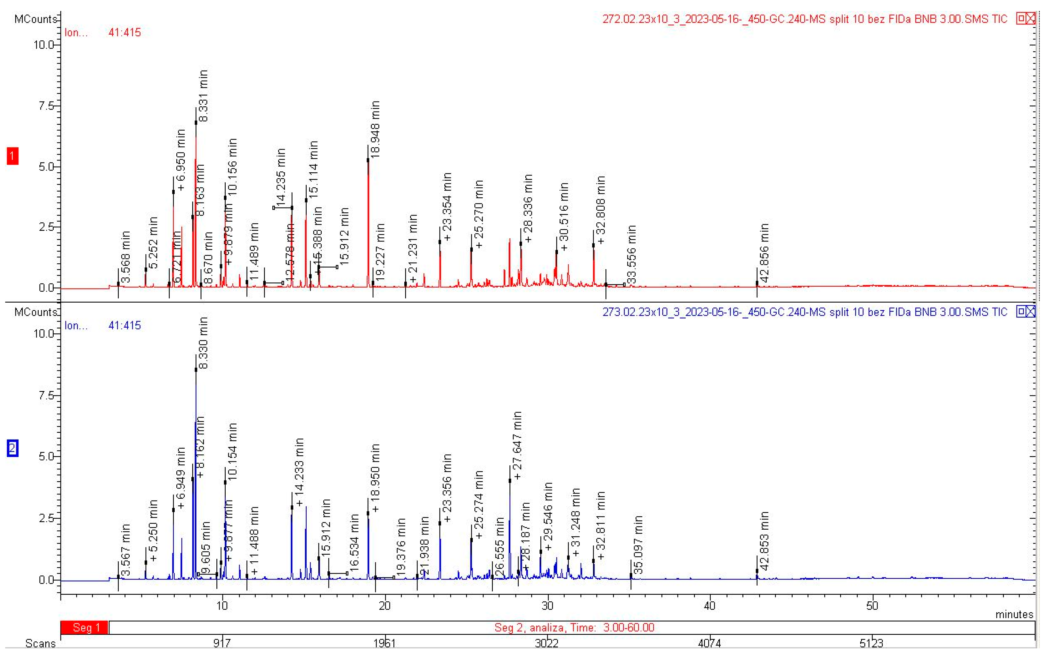
2.4. Analysis of the EO Composition

As shown in the present study, the dried herb contained 2.4 mL/kg of EO, whereas 2.5-fold greater amounts were obtained from the flowers, i.e., on average 6.1 mL/kg. These levels accounted for 0.235% and 0.598%, respectively. The EO extracted from the herb of Achillea millefolium subsp. millefolium contained 70 substances, which accounted for 96.24 relative percentages. In turn, 68 compounds (95.04 relative percentages) were detected in the EO from the flowers (Table 4, Figure 5).
β-pinene turned out to be the main component of both EOs, representing 9.90% and 12.22%, respectively, in the herb and flowers. Substantial amounts (over 5%) of 1,8 cineole (5.83% and 6.45%), camphor (5.81% and 5.28%), and borneol (6.18% and 5.25%) were detected in both EO samples. In terms of the qualitative composition, the EO extracted from the herb and flowers was similar, and there were substantial differences only in the amounts of individual components, especially bornyl acetate, accounting for 9.23% in the herb and 4.81% in the flowers, and α-pinene, whose content was 5.29% and 3.83%, respectively. In turn, the flower EO contained greater amounts of (E)-nerolidol (7.34% vs. 3.47% in the herb), sabinene (6.04% vs. 4.18% in the herb), and β-pinene (12.22 and 9.90%) (Table 4; Figure 5 and Figure 6).
The EO from the herb of Achillea millefolium subsp. millefolium had higher content of oxygenated compounds, mainly oxygenated monoterpenoids (25.58%), compared to the EO extracted from the inflorescences (20.88%). The herb also contained greater amounts of oxygenated sesquiterpenoids (6.56%) than the flowers (3.87%). Both EO samples had greater amounts of monoterpenes with derivatives than sesquiterpenes and their derivatives (Figure 7).

3. Discussion

3.1. Structure and Histochemistry

As in other Asteraceae representatives, the shoots of yarrow plants are composed of both exogenous and endogenous secretory tissues [51,52]. The exogenous secretory structures include biseriate glandular trichomes, which are typical of plants from the family Asteraceae. In the present study, the secretory trichomes in A. millefolium subsp. millefolium were located on the corollas of disc and ray flowers, receptacular bracts, and involuclar bracts. A similar distribution of trichomes on floral corollas and/or involuclar bracts in other Asteraceae representatives has been reported by Ferreira and Janick [61], Cornara et al. [62], Heinrich et al. [63], Sulborska [64,65], and Haratym et al. [53]. The glandular trichomes located on the stems, leaves, and flowers of A. millefolium subsp. millefolium were composed of three tiers of secretory cells. The present analyses revealed that the cells contained EO and substances typical of EO, i.e., neutral lipids and sesquiterpenes.
We also found these EO components in the epidermis cells of the corolla teeth and tubes of the small disc flowers. The apical zones of the adaxial surface of the teeth had conical cells (papillae), which contained EO as well. Conical cells located on flower petals frequently release scent [66]. The other functions of conical cells, which are most often found in the top region of petals, i.e., a site of direct contact with pollinators, are to increase their grip on the flower and decrease flower wettability [67,68,69].
The higher content of EO in A. millefolium subsp. millefolium flowers than in shoots (i.e., flowers, stems, and leaves) was associated with the presence of EO not only in the glandular trichomes located on the petals, receptacular, and involuclar bracts, but also in the petal epidermis cells (including papillae).

3.2. Quantity and Quality of EO

As indicated by the European Pharmacopoeia [18], whole or cut dried flowering tops of shoots containing at least 2 mL/kg of EO are the Achillea millefolium medicinal raw material. In the present study, the dried herb contained 2.4 mL/kg of EO, while the distillation yield from the inflorescences was on average 6.1 mL/kg; these values accounted for 0.235% and 0.598%, respectively. Other studies conducted in Poland reported highly diverse EO content ranging from 0.17 to 0.60% in A. millefolium herb originating from natural habitats [70]. Zawiślak and Nurzyńska-Wierdak [71] found that yarrow herb collected from wild plants had significantly higher amounts of EO (on average 0.5%) than herb of cultivated plants (on average 0.3%). Similarly, Benedek et al. [72] reported highly diverse levels of EO (from 0.5 to 5.88%) in commercial Achillea samples from Germany, Austria, France, Italy, the Netherlands, Great Britain and Bosnia-Herzegovina and in plants collected in the wild in Eastern European countries (Poland, Romania, Slovakia, Czechia, Hungary); however, the researchers did not focus on differences in the EO content between yarrow herb and flowers. In turn, Mohammed et al. [13] reported that the yield of EO from Achillea samples collected in Saudi Arabia ranged from 0.33% to 0.65%, with differences between the distillation yield from leaves, stems, and aerial parts of A. millefolium; the lowest yield was obtained from leaves. However, the authors did not specify the stage of plant development during collection or the presence and quantity of flowers in the collected aerial parts.
The EO from the inflorescences and herb of A. millefolium subsp. millefolium extracted in the present study contained the following main ingredients: β-pinene, bornyl acetate, (E)-nerolidol, 1,8-cineole, borneol, sabinene, camphor, and α-pinene. They were reported as the main components in many samples of yarrow raw material analysed in various countries: Norway (α-pinene, β-pinene, borneol, bornyl acetate, sabinene) [73], Estonia (β-pinene, 1,8-cineole, sabinene) [74], Iran (1,8-cineole, camphor) [28], and Lithuania ((E)-nerolidol, β-pinene and 1,8-cineole) [75]. However, we did not identify azulene or chamazulene, which were previously shown in many studies on Millefolii herba [9,28,57,76,77]. Azulenes, which are formed from proazulenes, are products of the dehydration of sesquiterpenes [78]. Likewise in the present study, various authors reported that yarrow EO did not contain chamazulene [13,79,80]. Orav et al. [56] found considerable differences in the composition of EO (19 samples) from A. millefolium growing in various European countries. They identified both chemotypes that were rich in chamazulene and chemotypes that did not contain this compound. In turn, Mockute and Judzentiene [81] identified four chemotypes of yarrow EO depending on the plant growth environment. Forty samples of EO were collected from plants growing in the wild in 21 localities of Lithuania; fourteen samples did not contain chamazulene, and two samples contained only traces of this component. The level of EO in 18 samples was in the range of 0.1–6.8%, and six samples contained 9.8–23.2% of EO. Based on these reports, the EOs analysed in the present study, which were characterised by the absence of chamazulene and the presence of β-pinene + 1,8-cineole, can be classified as chemotype IV in the classification proposed by Mockute and Judzentiene [81].
In the present study, the flowers of the analysed species contained higher levels of β-pinene and (E)-nerolidol than the herb. Inflorescence samples analysed by Judzentiene and Mockute [75] also contained substantially higher amounts of these compounds than leaves.
According to the requirements of the European Pharmacopoeia [18], the Achillea millefolium EO, which does not contain proazulenes, does not meet the requirements for use in traditional medicine. Although proazulenes are a measure of the quality of Achillea millefolium raw material for medical use, it should be emphasised that other EO components of this taxon also have great application-related importance. α- and β-Pinene and other dominant compounds have healing properties and are used in the cosmetic industry and for food flavouring [7,12,82,83,84,85,86].
The results of the present morpho-anatomical analyses and histochemical assays indicating the localisation of EO in the examined organs of A. millefolium subsp. millefolium correspond to the EO quantities revealed by phytochemical studies.

4. Materials and Methods

4.1. Plant Material

The materials for anatomical and phytochemical studies were Achillea millefolium subsp. millefolium L. capitula composed of ray and disc flowers (Millefolii flos) and upper parts of flowering shoots, i.e., herbal raw material (Millefolii herba). The research material was collected at the turn of July and August 2022 from natural meadows in Wola Zadybska near Lublin (the south-eastern Poland) (51°44′49″ N; 21°50′38″ E. The botanical identification of the plants was made by taxonomy specialist Professor Bożena Denisow. The collected plant specimens were also compared with authentic samples deposited in the Department of Botany and Plant Physiology, University of Life Sciences in Lublin (Poland) (no. 98).

4.2. Microscopic Preparation

4.2.1. Light Microscopy (LM)

Ten fresh capitula, from which disc (n = 20) and ray (n = 20) flowers were randomly sampled, were selected for microscopic observations. Fresh shoots in the full flowering phase (n = 10), from which 1 cm long stem fragments were cut off 0.5–2 cm below the inflorescence, and leaves (n = 10) were used as well. The preliminary observations were performed with the use of an STM 800 stereoscopic microscope (Microlab, Shenzhen, China), and photographs were taken for documentation.
Cross-sections of the stem and leaf were made by hand, and then microscopic slides were prepared in glycerine and water (1:1). Microscopic observations were performed using a Nikon Eclipse 400 light microscope (Nikon, Tokyo, Japan). The biseriate glandular trichomes located on the surface of the ray and disc flowers, receptacular bracts, involuclar bracts, leaves, and stems were subjected to morphometric measurements. Their length and width were measured at the widest point (n = 30 each). The length and width of non-glandular trichomes located on the stems, leaves, receptacular bracts, and involuclar bracts were measured as well (n = 30 from each organ).
Semi-thin sections of fresh stems and leaves were prepared. One-centimetre-long stem fragments were taken from the middle part of the third internode (counting from the inflorescences). One-centimetre-long fragments of the leaf blade with the midrib were collected from the apical part. The plant material was fixed in 2.5% glutaraldehyde in phosphate buffer (pH 7.2; 0.1 M) at 4 °C for 12 h. In the next step, the samples were washed three times in phosphate buffer, dehydrated in an ethanol series, and embedded in LR white resin (LR white acrylic resin, medium grade, Sigma-Aldrich, Saint-Louis, MO, USA). Semi-thin sections (0.7–0.9 μm) were cut transversally using a Reichert Ultracut S ultramicrotome (C. Reichert Optische Werke AG, Vienne, Austria) and a glass knife. For general histology, the sections were stained with a 1% (w/v) aqueous methylene blue-azure II solution [87]. The sections were examined using an Olympus CX 23 light microscope (Olympus, Tokyo, Japan) equipped with an Olympus EP50 digital camera (Olympus, Tokyo, Japan) and EPview software ver. 1.2.

4.2.2. Scanning Electron Microscopy (SEM)

The surfaces of the stems, leaves, and flowers were observed using SEM. Stem and leaf fragments 0.5 cm long (n = 10) were fixed in a 2.5% glutaraldehyde solution in 0.1 M phosphate buffer (pH 7.0) at room temperature for 2 h and then washed in phosphate buffer four times at 20-min intervals. The fixed plant material was dehydrated in graded ethanol series and immersed in absolute ethanol (POCH, Gliwice, Poland) three times for 30 min. Next, the flower samples were critical-point dried in liquid CO2 using a K 850 Critical Point Dryer and sputter-coated with gold (20 mm thickness) using a K 550X device (Emitech, Ashford, UK). The observations were made with the use of a Tescan Vega II LMU scanning electron microscope (Tescan, Brno, Czech Republic) at an accelerating voltage of 30 kV. Images were taken using a BSE detector and saved using Vega software ver. 3.5.19.

4.3. Histochemical Assays

Disc flowers, receptacular bracts (n = 20 each), and hand-made cross-sections of the stems (0.5–2 cm below the inflorescence; n = 10) were subjected to histochemical analyses (n = 6) to identify lipophilic compounds present in these organs and determine their location. Sudan IV [88,89] and Neutral red [90,91] staining was employed to identify total lipids and EO, Nile blue was used to detect acidic and neutral lipids [92,93], and sesquiterpenes were detected using concentrated sulphuric acids [94,95]. The flowers and stem sections were also used to make control aqueous glycerol preparations (1:1). All preparations were observed under an Olympus CX23 light microscope (Olympus, Tokyo, Japan) equipped with a digital camera Olympus EP50 (Olympus, Tokyo, Japan) and EPview software ver. 1.2.

4.4. Phytochemical Analysis

Fully blooming yarrow shoots were cut at a height of approximately 20 cm above the ground. Immediately after harvesting, the material was dried in a dryer with forced air circulation at a temperature of 35 °C. The samples were dried for 2 h to achieve a constant humidity level of 12%. The dried material was mixed and homogenised to obtain herb containing flowers, leaves, and thin fragments of stems, whereas fully blooming inflorescences were separated (Figure 8). In the study, we used 500 g of dry herb material and 300 g of floral material. Fifty-gram samples of the plant material were hydrodistilled in a Deryng apparatus for 3 h. The distillation process was repeated several times to obtain at least 1 mL of EO from each sample type. We obtained 1.8 mL of floral EO and 1.2 mL of herb EO from the collected material. The content of EO in the raw material was expressed in mL/kg. The EO samples were diluted 1000-fold in hexane. The analysis of the EO composition was carried out using a gas chromatograph with a 450-GC + 240-MS Varian mass detector at the Central Research Laboratory of the University of Life Sciences in Lublin.
The following conditions were used: sample dose: 1 µL, dispenser: 250 °C, split 1:10, carrier gas: helium, constant flow 1.0 mL/min, column: VF-5ms (DB-5 equivalent), column oven: 50 °C held for 1 min, ramp to 240 °C at a rate of 4 °C/min, 240 °C held for 10 min, total analysis time: 60 min, detector: mass spectrometer with an ion trap: electron ionisation EI, full scanning: in the range of 41–415 m/z, scanning speed: 0.8 s/scan. The compounds were identified based on mass spectra and “Identification of Essential Oil Components by Gas Chromatography/Mass Spectrometry, ed. 4.1; Dr. Robert P. Adams Professor Biology Department Baylor University” and the NIST Mass Spectra Library.
Kovats retention indices were determined on the VF-5ms column (equivalent to DB-5) using a series of n-alkanes (C7-C40 Saturated Alkanes Standard, Product no.: 4 9452-U Supelco) as in Van Den Dool and Kratz [96].

5. Conclusions

The stems, leaves, and flowers of Achillea millefolium subsp. millefolium, which are medicinal and cosmetic raw material, were found to have exo- and endogenous secretory structures responsible for the production and accumulation of EO components. The presence of EO was detected in glandular trichomes, papillae, and secretory canals. In turn, sesquiterpenes, which are important components of EO as well, were detected not only in glandular trichomes but also in disc flower epidermis. The analysis of the distillation yield from the herb samples (2.4 mL/kg) and flowers (6.1 mL/kg) of A. millefolium subsp. millefolium showed that the flowers contained substantially higher amounts of EO than the other parts of the shoot. The composition of the EO extracted from both samples was largely similar, with higher content of monoterpenes and their derivatives than sesquiterpenes and their derivatives. In turn, the herb EO exhibited a higher number of oxygenated monoterpenes and oxygenated sesquiterpenoids than the EO obtained from the flowers. The analysed A. millefolium subsp. millefolium plants represented the azulene-free type. The EOs extracted from both the herb and flowers represent the β-pinene + 1,8-cineole chemotype. It can be expected that, given the biological and medicinal activity of the dominant EO components, the analysed yarrow chemotype can be used in the pharmaceutical and cosmetic industries and for food flavouring. As shown in this study, the presence of various secretory structures in the flower epidermis (more numerous glandular trichomes and papillae) contributed to the higher amounts of EO produced by the flowers than by the herb.

Author Contributions

Conceptualization, E.W.-C.; Methodology, A.K., E.W.-C., A.K.-D., A.S.-R., M.D. and M.G. Formal analysis, E.W.-C., A.S.-R., A.K., A.K.-D., M.G. and M.D., Investigation, A.K., A.S.-R., A.K.-D. and M.D.; Writing—original draft preparation, A.K., E.W.-C., A.K-D., A.S.-R., M.G. and M.D.; Visualization, A.S.-R., A.K. and A.K.-D.; Funding acquisition, A.K. and A.S.-R. All authors have read and agreed to the published version of the manuscript.

Funding

This research was funded by Poland’s Ministry of Education and Science as part of the statutory activities of the Department of Botany and Plant Physiology, University of Life Sciences in Lublin (project LKR/S/49/2023RiO).

Institutional Review Board Statement

Not applicable.

Informed Consent Statement

Not applicable.

Data Availability Statement

The datasets used and/or analysed during the current study are available from the corresponding author on reasonable request.

Conflicts of Interest

The authors declare no conflict of interest.

References

  1. Ehrendorfer, F.; Guo, Y.P. Multidisciplinary studies on Achillea sensu lato (Compositae-Anthemideae): New data on systematics and phylogeography. Willdenowia 2006, 36, 69–87. [Google Scholar] [CrossRef]
  2. Zengin, G.; Aktumsek, A.; Ceylan, R.; Uysal, S.; Mocan, A.; Guler, G.O.; Mahomoodally, M.F.; Glamočlija, J.; Ćirićf, A.; Soković, M. Shedding light on the biological and chemical fingerprints of three Achillea species (A. biebersteinii, A. millefolium and A. teretifolia). Food Funct. 2017, 8, 1152–1165. [Google Scholar] [CrossRef]
  3. Afshari, M.; Rahimmalek, M.; Miroliaei, M. Variation in polyphenolic profiles, antioxidant and antimicrobial activity of different Achillea species as natural sources of antiglycative compounds. Chem. Biodivers. 2018, 15, e1800075. [Google Scholar] [CrossRef] [PubMed]
  4. Salehi, B.; Selamoglu, Z.; Sevindik, M.; Fahmy, N.M.; Al-Sayed, E.; El-Shazly, M.; Büsselberg, D. Achillea spp.: A comprehensive review on its ethnobotany, phytochemistry, phytopharmacology and industrial applications. Cell. Mol. Biol. 2020, 66, 78–103. [Google Scholar] [CrossRef] [PubMed]
  5. Barda, C.; Grafakou, M.E.; Tomou, E.M.; Skaltsa, H. Phytochemistry and evidence-based traditional uses of the genus Achillea L.: An update (2011–2021). Sci. Pharm. 2021, 89, 50. [Google Scholar] [CrossRef]
  6. Applequist, W.L.; Moerman, D.E. Yarrow (Achillea millefolium L.): A neglected panacea? A review of ethnobotany, bioactivity, and biomedical research. Econ. Bot. 2011, 65, 209–225. [Google Scholar] [CrossRef]
  7. Kohlmünzer, S. Farmakognozja; PZWL Wydawnictwo Lekarskie: Warszawa, Poland, 2020; p. 599. [Google Scholar]
  8. Guo, Y.P.; Saukel, J.; Ehrendorfer, F. AFLP trees versus scatterplots: Evolution and phylogeography of the polyploid complex Achillea millefolium agg. (Asteraceae). Taxon 2008, 57, 153–169. [Google Scholar] [CrossRef]
  9. Bączek, K.; Kosakowska, O.; Przybył, J.L.; Kuźma, P.; Ejdys, M.; Obiedziński, M.; Węglarz, Z. Intraspecific variability of yarrow (Achillea millefolium L. s.l.) in respect of developmental and chemical traits. Herba Pol. 2015, 61, 37–52. [Google Scholar] [CrossRef]
  10. Štrbac, F.; Bosco, A.; Amadesi, A.; Rinaldi, L.; Stojanović, D.; Simin, N.; Orčić, D.; Pušić, I.; Krnjajić, S.; Ratajac, R. In Vitro Ovicidal Activity of Two Chemotypes of the Yarrow (Achillea millefolium L.) Essential oil against ovine gastrointestinal nematode eggs. Arhiv Vet. Med. 2020, 13, 59–76. [Google Scholar] [CrossRef]
  11. The Plant List. Available online: www.theplantlist.org (accessed on 25 August 2023).
  12. Nowak, K.; Ogonowski, J.; Szulc, K. Application and characteristics of Achillea millefolium and its oil. Chemik 2010, 64, 103–110. [Google Scholar]
  13. Mohammed, H.A.; Abd-Elraouf, M.; Sulaiman, G.M.; Almahmoud, S.A.; Hamada, F.A.; Khan, R.A.; Hegazy, M.M.; Abd-El-Wahab, M.F.; Kedra, T.A.; Ismail, A. Variability in the volatile constituents and biological activities of Achillea millefolium L. essential oils obtained from different plant parts and by different solvents. Arab. J. Chem. 2023, 16, 105103. [Google Scholar] [CrossRef]
  14. Abou Baker, D.H. Achillea millefolium L. ethyl acetate fraction induces apoptosis and cell cycle arrest in human cervical cancer (HeLa) cells. Ann. Agric. Sci. 2020, 65, 42–48. [Google Scholar] [CrossRef]
  15. Gaweł-Bęben, K.; Strzępek-Gomółka, M.; Czop, M.; Sakipova, Z.; Głowniak, K.; Kukula-Koch, W. Achillea millefolium L. and Achillea biebersteinii Afan. hydroglycolic extracts–bioactive ingredients for cosmetic use. Molecules 2020, 25, 3368. [Google Scholar] [CrossRef]
  16. Strzępek-Gomółka, M.; Gaweł-Bęben, K.; Kukula-Koch, W. Achillea species as sources of active phytochemicals for dermatological and cosmetic applications. Oxid. Med. Cell. Longev. 2021, 3, 6643827. [Google Scholar] [CrossRef] [PubMed]
  17. Łuczaj, Ł.; Pieroni, A.; Tardío, J.; Pardo-de-Santayana, M.; Sõukand, R.; Svanberg, I.; Kalle, R. Wild food plant use in 21st century Europe: The disappearance of old traditions and the search for new cuisines involving wild edibles. Acta Soc. Bot. Pol. 2012, 81, 359–370. [Google Scholar] [CrossRef]
  18. European Pharmacopoeia 11. Yarrow. Millefolii herba. 2023. Available online: https://www.edqm.eu/en/european-pharmacopoeia-ph.-eur.-11th-edition (accessed on 16 June 2023).
  19. Daniel, P.S.; Lourenço, E.L.B.; Sete da Cruz, R.M.; de Souza Goncalves, C.H.; Marques Das Almas, L.R.; Hoscheid, J.; da Silva, C.; Jacomass, E.; Junior, R.B.; Alberton, O. Composition and antimicrobial activity of essential oil of yarrow (Achillea millefolium L.). Aust. J. Crop Sci. 2020, 14, 545–550. [Google Scholar] [CrossRef]
  20. Askari, F.; Mirza, M.; Tafti, M.M. Effect of density and year of cultivation on essential oil content and chemical compounds of Achillea millefolium subsp. elbursensis. Indian J. Nat. Prod. Resour. 2021, 12, 122–127. [Google Scholar]
  21. Boris, A.J.; Takahama, S.; Weakley, A.T.; Debus, B.M.; Shaw, S.L.; Edgerton, E.S.; Joo, T.; Ng, N.L.; Dillner, A.M. Quantifying organic matter and functional groups in particulate matter filter samples from the southeastern United States—Part 2: Spatiotemporal trends. Atmos. Meas. Tech. 2021, 14, 4355–4374. [Google Scholar] [CrossRef]
  22. Ni, Z.; Wu, Y.; Zhang, K.; Dong, M.; Sauriol, F.; Huo, C.; Gu, Y.; Shi, Q. A monoterpene and two sesquiterpenoids from the flowers of Achillea millefolium. Chem. Nat. Compd. 2013, 49, 450–453. [Google Scholar] [CrossRef]
  23. Arias-Durán, L.; Estrada-Soto, S.; Hernández-Morales, M.; Millán-Pacheco, C.; Navarrete-Vázquez, G.; Villalobos-Molina, R.; Ibarra-Barajas, M.; Almanza-Pérez, J.C. Antihypertensive and vasorelaxant effect of leucodin and achillin isolated from Achillea millefolium through calcium channel blockade and NO production: In vivo, functional ex vivo and in silico studies. J. Ethnopharmacol. 2021, 273, 113948. [Google Scholar] [CrossRef]
  24. Leino, T.O.; Sieger, P.; Yli-Kauhaluoma, J.; Wallén, E.A.A.; Kley, J.T. The azulene scaffold from a medicinal chemist’s perspective: Physicochemical and in vitro parameters relevant for drug discovery. Eur. J. Med. Chem. 2022, 237, 114374. [Google Scholar] [CrossRef] [PubMed]
  25. Villanueva-Bermejo, D.; Zahran, F.; Troconis, D.; Villalva, M.; Reglero, G.; Fornari, T. Selective precipitation of phenolic compounds from Achillea millefolium L. extracts by supercritical anti-solvent technique. J. Supercrit. Fluids 2017, 120, 52–58. [Google Scholar] [CrossRef]
  26. Salomon, L.; Lorenz, P.; Bunse, M.; Spring, O.; Stintzing, F.C.; Kammerer, D.R. 2021 Comparison of the Phenolic Compound Profile and Antioxidant Potential of Achillea atrata L. and Achillea millefolium L. Molecules 2021, 26, 1530. [Google Scholar] [CrossRef] [PubMed]
  27. Edreva, A.; Vitkova, A.; Gesheva, E. Field-Cultivated plants from Achillea millefolium group: Total flavonoid content, antiradical and antioxidant activities in stems and leaves, and ratio of plant parts. Genet. Plant Physiol. 2019, 9, 3–10. [Google Scholar]
  28. Farhadi, N.; Babaei, K.; Farsaraei, S.; Moghaddam, M.; Pirbaloti, A.G. Changes in essential oil compositions, total phenol, flavonoids and antioxidant capacity of Achillea millefolium at different growth stages. Ind. Crops Prod. 2020, 152, 112570–112576. [Google Scholar] [CrossRef]
  29. Grigore, A.; Colceru-Mihul, S.; Bazdoaca, C.; Yuksel, R.; Ionita, C.; Glava, L. Antimicrobial activity of an Achillea millefolium L. Proceedings 2020, 57, 34. [Google Scholar] [CrossRef]
  30. Adil, M.; Dastagir, G.; Bakht, J.; Ambrin, A. Phytochemical screening and antimicrobial activity of medicinally important Achillea millefolium L. and Chaerophyllum villosum Wall Exdc. Pak. J. Bot. 2020, 52, 971–974. [Google Scholar] [CrossRef]
  31. Karimi, A.; Niazkar, H.R.; Azar, P.S.; Tutunchi, H.; Karimi, M.; Asghariazar, V.; Kooshki, F. Protective effect of hydro-alcoholic extract of Achillea millefolium on renal injury and biochemical factors in streptozotocin-induced diabetic rats. Nutr. Food Sci. 2021, 51, 1068–1083. [Google Scholar] [CrossRef]
  32. Jahromi, G.P.; Imani, E.; Nasehi, M.; Shahriari, A. Effect of Achillea millefolium aqueous extract on memory deficit and anxiety caused by stroke in ovariectomized rats. J. Herbmed Pharmacol. 2019, 8, 153–159. [Google Scholar] [CrossRef]
  33. Varzaneh, F.E.; Nahidi, F.; Mojab, F.; Pourhoseingholi, M.A. Effect of Achillea millefolium on the intensity and duration of menstrual bleeding of women with menorrhagia. Iran. J. Obstet. Gynecol. Infertil. 2020, 23, 67–77. [Google Scholar] [CrossRef]
  34. Niazipoor, G.; AghaAlikhani, M.; Mokhtassi-Bidgoli, A.; Vitalini, S. Essential oil profile of yarrow (Achillea spp. and Tanacetum spp.) ecotypes and their allelopathic potential to suppress redroot pigweed (Amaranthus retroflexus L.). Res. Sq. 2023, in press. [Google Scholar] [CrossRef]
  35. Alomair, M.K.; Alabduladheem, L.S.; Almajed, M.A.; Alobaid, A.A.; Alkhalifah, E.A.; Younis, N.S.; Mohamed, M.E. Achillea millefolium essential oil mitigates peptic ulcer in rats through Nrf2/HO-1 pathway. Molecules 2022, 27, 7908. [Google Scholar] [CrossRef]
  36. Alzomor, A.K.Y.; Al-Absi, N.H.; Al-hssany, A.F.; Al-Salahi, H.S.; Almushra’a, A.A. Investigate the effects of Achillea millefolium plant extract as a hepatoprotection on carbon tetrachloride-induced liver toxicity in female rats. Saudi J. Med. Pharm. Sci. 2022, 8, 227–233. [Google Scholar] [CrossRef]
  37. Becker, L.C.; Bergfeld, W.F.; Belsito, D.V.; Hill, R.A.; Klaassen, C.D.; Liebler, D.C.; Marks, M.G., Jr.; Shank, R.C.; Slaga, T.J.; Snyder, P.W.; et al. Safety assessment of Achillea millefolium as used in cosmetics. Int. J. Toxicol. 2016, 35 (Suppl. S3), 5S–15S. [Google Scholar] [CrossRef] [PubMed]
  38. Calapai, G.; Casciaro, M.; Miroddi, M.; Calapai, F.; Navarra, M.; Gangemi, S. Montelukast-induced adverse drug reactions: A review of case reports in the literature. Pharmacology 2014, 94, 60–70. [Google Scholar] [CrossRef]
  39. Hamzic, A.; Ginko, E.; Delic, N.; Kozarevic, E.C.; Izic, B.; Hamzic, D.; Sarić-Kundalic, B. Therapeutic effect of ointment for psoriasis based on Achillea millefolium L., Calendula officinalis L. and Salvia officinalis L. Technol. Acta 2022, 15, 69–79. [Google Scholar]
  40. Syakri, S.; Ismail, I.; Amal, N.M.; Masjidi, N.A.; Tahir, K.A. Characterization and anti-aging tests of peel-off gel masks made from ethanolic extract of yarrow (Achillea millefolium). Open Access Maced. J. Med. Sci. 2021, 9, 1156–1161. [Google Scholar] [CrossRef]
  41. CosIng Cosmetic Ingredient Database. Available online: https://ec.europa.eu/growth/sectors/cosmetics/cosing_en (accessed on 15 July 2023).
  42. Mistry, N. Guidelines for formulating anti-pollution products. Cosmetics 2017, 4, 57. [Google Scholar] [CrossRef]
  43. Tadić, V.; Arsić, I.; Zvezdanović, J.; Zugić, A.; Cvetković, D.; Pavkov, S. The estimation of the traditionally used yarrow (Achillea millefolium L. Asteraceae) oil extracts with anti-inflamatory potential in topical application. J. Ethnopharmacol. 2017, 199, 138–148. [Google Scholar] [CrossRef]
  44. Łuczaj, Ł. Changes in Assumption Day Herbal Bouquets in Poland: A nineteenth century study revisited. Econ. Bot. 2011, 65, 66–75. [Google Scholar] [CrossRef]
  45. Mohammadhosseini, M.; Sarker, S.D.; Akbarzadeh, A. Chemical composition of the essential oils and extracts of Achillea species and their biological activities: A review. J. Ethnopharmacol. 2017, 199, 257–315. [Google Scholar] [CrossRef] [PubMed]
  46. Kalle, R.; Sõukand, R.; Pieroni, A. Devil is in the details: Use of wild food plants in historical Võromaa and Setomaa, present-day Estonia. Foods 2020, 9, 570. [Google Scholar] [CrossRef] [PubMed]
  47. Darnahal, E.; Jamshidi, M.; Jafarlou, M.; Hasheminia, S. Insecticidal and repellent effects of essential oilsfrom different parts of Achillea millefolium against adult of Oryzaephilus surinamensis L. (Coleoptera, Silvanidae). Appl. Plant Prot. 2018, 7. in press. [Google Scholar]
  48. Figueiredo, A.C.; Pais, M.S.S. Ultrastructural aspects of the glandular cells from the secretory trichomes and from the cell suspension cultures of Achillea millefolium L. ssp. millefolium. Ann. Bot. 1994, 74, 179–190. [Google Scholar] [CrossRef]
  49. Aytas Akcin, T.; Akcin, A. Morphological and anatomical characteristics and taxonomical significance of achene micromorphology of Achillea phrygia and A. gypsicola (Asteraceae), endemic to Turkey. Nord. J. Bot. 2010, 28, 65–73. [Google Scholar] [CrossRef]
  50. Afshari, M.; Rahimmalek, M. Variation in essential oil composition, bioactive compounds, anatomical and antioxidant activity of Achillea aucheri, an endemic species of Iran, at different phenological stages. Chem. Biodivers. 2018, 15, e1800319. [Google Scholar] [CrossRef] [PubMed]
  51. Ilham, M.; Mukarromah, S.R.; Rakashiwi, G.A.; Indriati, D.I.; Yoku, B.F.; Purnama, P.R.; Junairiah, J.; Prasongsuk, S.; Purnobasuki, H.; Wahyuni, D.K. Morpho-anatomical characterization and DNA barcoding of Achillea millefolium L. Biodiversitas 2022, 23, 1958–1969. [Google Scholar] [CrossRef]
  52. Tekin, M.; Akdere, Ş. Anatomical investigations of the Turkish critically endangered species: Achillea sivasica Çelik et Akpulat (Asteraceae). Acta Bot. Croat. 2021, 80, 91–98. [Google Scholar] [CrossRef]
  53. Haratym, W.; Weryszko-Chmielewska, E.; Konarska, A. Microstructural and histochemical analysis of aboveground organs of Centaurea cyanus used in herbal medicine. Protoplasma 2020, 257, 285–298. [Google Scholar] [CrossRef]
  54. Mercado, M.I.; Marcial, G.; Catalán, J.V.; Grau, A.; Catalán, C.A.; Ponessa, G.I. Morphoanatomy, histochemistry, essential oil, and other secondary metabolites of Artemisia copa (Asteraceae). Bot. Lett. 2021, 168, 577–593. [Google Scholar] [CrossRef]
  55. Paes de Almeida, V.; Heiden, G.; Raman, V.; Novatski, A.; Bussade, J.E.; Farago, P.V.; Manfron, J. Microscopy and histochemistry of leaves and stems of Baccharis subgenus Coridifoliae (Asteraceae) through LM and SEM-EDS. Microsc. Microanal. 2021, 27, 1273–1289. [Google Scholar] [CrossRef]
  56. Orav, A.; Arak, E.; Raal, A. Phytochemical analysis of the essential oil of Achillea millefolium L. from various European Countries. Nat. Prod. Res. 2006, 20, 1082–1088. [Google Scholar] [CrossRef] [PubMed]
  57. Mazandarani, M.; Mirdeilami, S.Z.; Pessarakli, M. Essential oil composition and antibacterial activity of Achillea millefolium L. from different regions in North east of Iran. J. Med. Plants Res. 2013, 7, 1063–1069. [Google Scholar]
  58. Aćimović, M.; Zorić, M.; Zheljazkov, V.D.; Pezo, L.; Čabarkapa, I.; Stanković Jeremić, J.; Cvetković, M. Chemical characterization and antibacterial activity of essential oil of medicinal plants from Eastern Serbia. Molecules 2020, 25, 5482. [Google Scholar] [CrossRef]
  59. Turk, B.; Baričevič, D.; Batič, F. Essential oil content, chamazulene content and antioxidative properties of Achillea millefolium agg. extracts from Slovenia. Acta Agric. Slov. 2021, 117, 1–10. [Google Scholar] [CrossRef]
  60. Garzoli, S.; Cicaloni, V.; Salvini, L.; Trespidi, G.; Iriti, M.; Vitalini, S. SPME-GC-MS Analysis of the volatile profile of three fresh yarrow (Achillea millefolium L.) Morphotypes from different regions of Northern Italy. Separations 2023, 10, 51. [Google Scholar] [CrossRef]
  61. Ferreira, J.F.S.; Janick, J. Floral morphology of Artemisia annua with special reference to trichomes. Int. J. Plant. Sci. 1995, 156, 807–815. [Google Scholar]
  62. Cornara, L.; Bononi, M.; Tateo, F.; Serrato-Valenti, G.; Mariotti, M.G. Trichomes on vegetative and reproductive organs of Stevia rebaudiana (Asteraceae). Structure and secretory products. Plant Biosyst. 2001, 135, 25–37. [Google Scholar] [CrossRef]
  63. Heinrich, G.; Pfeifhofe, H.W.; Stabentheiner, E.; Sawidis, T. Glandular hairs Sigesbeckia jorullensis Kunth (Asteraceae): Morphology, histochemistry and composition of essential oil. Ann. Bot. 2002, 89, 459–469. [Google Scholar] [CrossRef]
  64. Sulborska, A. Micromorphology of flowers, anatomy and ultrastructure of Chamomilla recutita (L.) Rausch. (Asteraceae) nectary. Acta Agrobot. 2011, 64, 23–34. [Google Scholar] [CrossRef]
  65. Sulborska, A. Structure and distribution of glandular and non-glandular trichomes on above-ground organs in Inula helenium L. (Asteraceae). Acta Agrobot. 2013, 66, 25–34. [Google Scholar] [CrossRef]
  66. Reed, A.; Rudall, P.J.; Brockington, S.F.; Glover, B.J. Conical petal epidermal cells, regulated by the MYB transcription factor MIXTA, have an ancient origin within the angiosperms. J. Exp. Bot. 2022, 73, 5490–5502. [Google Scholar] [CrossRef] [PubMed]
  67. Ojeda, D.I.; Valido, A.; Fernández de Castro, A.G.; Ortega-Olivencia, A.; Fuertes-Aguilar, J.; Carvalho, J.A.; Santos-Guerra, A. Pollinator shifts drive petal epidermal evolution on the Macaronesian Islands bird-flowered species. Biol. Lett. 2016, 12, 20160022. [Google Scholar] [CrossRef] [PubMed]
  68. Bailes, E.J.; Glover, B.J. Intraspecific variation in the petal epidermal cell morphology of Vicia faba L. (Fabaceae). Flora 2018, 244–245, 29–36. [Google Scholar] [CrossRef] [PubMed]
  69. Stavenga, D.G.; Staal, M.; Van der Kooi, C.J. Conical epidermal cells cause velvety coloration and enhanced patterning in Mandevilla flowers. Faraday Discuss. 2020, 223, 98–106. [Google Scholar] [CrossRef]
  70. Bączek, K.; Kosakowska, O.; Przybył, J.; Kuczerenko, A.; Pióro-Jabrucka, E.; Węglarz, Z. Zróżnicowanie chemiczne dziko rosnących populacji krwawnika pospolitego (Achillea millefolium L.). J. Agronom. 2013, 15, 89–94. [Google Scholar]
  71. Zawiślak, G.; Nurzyńska-Wierdak, R. Plon surowca uprawianych oraz dziko rosnących roślin krwawnika pospolitego (Achillea millefolium L.) i wrotyczu pospolitego (Tanacetum vulgare L.). Ann. Horti. 2017, 27, 27–35. [Google Scholar] [CrossRef]
  72. Benedek, B.; Rothwangl-Wiltschnigg, K.; Rozema, E.; Gjoncaj, N.; Reznicek, G.; Jurenitsch, J.; Kopp, B.; Glasl, S. Yarrow (Achillea millefolium L. sl): Pharmaceutical quality of commercial samples. Pharmazie 2008, 63, 23–26. [Google Scholar] [CrossRef]
  73. Rohloff, J.; Skagen, E.B.; Steen, A.H.; Iversen, T.H. Production of yarrow (Achillea millefolium L.) in Norway: Essential oil content and quality. J. Agric. Food Chem. 2000, 48, 6205–62059. [Google Scholar] [CrossRef]
  74. Orav, A.; Kailas, T.; Ivask, K. Composition of the essential oil from Achillea millefolium L. from Estonia. J. Essent. Oil Res. 2001, 13, 290–294. [Google Scholar] [CrossRef]
  75. Judzentiene, A.; Mockute, D. Essential oil composition of two yarrow taxonomic forms. Cent. Eur. J. Biol. 2010, 5, 346–352. [Google Scholar] [CrossRef]
  76. Zawiślak, G.; Nurzyńska-Wierdak, R. Rozwój roślin oraz skład chemiczny olejku eterycznego krwawnika pospolitego (Achillea millefolium L.) uprawianego w warunkach klimatu umiarkowanego. Ann. Horti. 2017, 27, 31–38. [Google Scholar] [CrossRef]
  77. Guz, L.; Wawrzykowski, J.; Adaszek, Ł. Anti-babesial potential and chemical composition of essential oil from yarrow Achillea millefolium. Pol. J. Vet. Sci. 2021, 24, 79–84. [Google Scholar] [CrossRef]
  78. Dąbrowska, J. 2023. Rozmieszczenie azulenowych i bezazulenowych form Achillea L. w Czechach. Herbalism 2023, 8, 152–165. [Google Scholar] [CrossRef]
  79. Candan, F.; Unlu, M.; Tepe, B.; Daferera, D.; Polissiou, M.; Sökmen, A.; Akpulat, H.A. Antioxidant and antimicrobial activity of the essential oil and methanol extracts of Achillea millefolium subsp. millefolium Afan. (Asteraceae). J. Ethnopharmacol. 2003, 87, 215–220. [Google Scholar] [CrossRef] [PubMed]
  80. Azizi, M.; Chizzola, R.; Ghani, A.; Oroojalian, F. Composition at different development stages of the essential oil of four Achillea species grown in Iran. Nat. Prod. Commun. 2010, 5, 283–290. [Google Scholar] [CrossRef]
  81. Mockute, D.; Judzentiene, A. Variability of the essential oils composition of Achillea millefolium ssp. millefolium growing wild in Lithuania. Biochem. Syst. Ecol. 2003, 31, 1033–1045. [Google Scholar] [CrossRef]
  82. Chan, W.-K.; Tan, L.T.-H.; Chan, K.-G., 3rd; Lee, L.-H.; Go, B.-H. (E)-nerolidol: A sesquiterpene alcohol with multi-faceted pharmacological and biological activities. Molecules 2016, 21, 529. [Google Scholar] [CrossRef]
  83. Vespermann, K.A.C.; Paulino, B.N.; Barcelos, M.C.S.; Pessôa, M.G.; Pastore, G.M.; Molina, G. Biotransformation of α- and β-pinene into flavor compounds. Appl. Microbiol. Biotechnol. 2017, 101, 1805–1817. [Google Scholar] [CrossRef]
  84. Arunachalam, S.; Nagoor Meeran, M.F.; Azimullah, S.; Sharma, C.; Goyal, S.N.; Ojha, S. (E)-nerolidol attenuates oxidative stress, inflammation, and apoptosis by modulating Nrf2/MAPK signaling pathways in doxorubicin-induced acute cardiotoxicity in rats. Antioxidants 2021, 10, 984. [Google Scholar] [CrossRef]
  85. Nyamwihura, R.J.; Ogungbe, I.V. The pinene scaffold: Its occurrence, chemistry, synthetic utility, and pharmacological importance. RSC Adv. 2022, 12, 11346–11375. [Google Scholar] [CrossRef] [PubMed]
  86. Hoch, C.C.; Petry, J.; Griesbaum, L.; Weiser, T.; Werner, K.; Ploch, M.; Verschoor, A.; Multhoff, G.; Dezfouli, A.B.; Wollenberg, B. 1,8-cineole (eucalyptol): A versatile phytochemical with therapeutic applications across multiple diseases. Biomed. Pharmacother. 2023, 167, 115467. [Google Scholar] [CrossRef] [PubMed]
  87. O’Brien, T.P.; McCully, M.E. The Study of Plant Structure: Principles and Selected Methods; Termarcarphi Pty. Ltd.: Melbourne, Australia, 1981. [Google Scholar]
  88. Johansen, D.A. Plant Microtechnique, 1st ed.; London McGraw Hill: London, UK, 1940. [Google Scholar]
  89. Lison, L. Histochimie et Cytochimie Animales, 3rd ed.; Gauthier-Villars: Paris, France, 1960. [Google Scholar]
  90. Kirk, P.W. Neutral red as a lipid fluorochrome. Stain Technol. 1970, 45, 1–4. [Google Scholar] [CrossRef] [PubMed]
  91. Conn, H.J. Biological Stains: A Handbook on the Nature and Uses of the Dyes Employed in the Biological Laboratory, 9th ed.; Williams & Wilkins: Baltimore, MD, USA, 1977. [Google Scholar]
  92. Cain, A.J. The use of Nile blue in the examination of lipids. Q. J. Microsc. Sci. 1947, 88, 383–392. [Google Scholar]
  93. Jensen, W.A. Botanical Histochemistry Principles and Practice, 1st ed.; WH Freeman and Company: San Francisco, CA, USA, 1962. [Google Scholar]
  94. Geissman, T.A.; Griffin, T.S. Sesquiterpene lactones: Acid-catalysed color reactions as an aid in structure determination. Phytochemistry 1971, 10, 2475–2485. [Google Scholar] [CrossRef]
  95. Cappelletti, E.M.; Caniato, R.; Appendino, G. Localization of the cytotoxic hydroperoxyeudesmanolides in Artemisia umbelliformis. Biochem. Syst. Ecol. 1986, 14, 183–190. [Google Scholar] [CrossRef]
  96. Van Den Dool, H.; Kratz, P.D. A generalization of the retention index system including linear temperature programmed gas-liquid partition chromatography. J. Chromatogr. 1963, 11, 463–471. [Google Scholar] [CrossRef]
Figure 1. Morphology of Achillea millefolium subsp. millefolium inflorescences and flowers; (ag,jl)—stereoscopic microscopy (SM), (h,i,mo)—light microscopy (LM), (p)—scanning electron microscopy (SEM). (a) Fragment of the upper stem part with capitula in the full-bloom stage; (b) General view of a capitula; white arrowheads indicate ray flowers, black arrowheads indicate disc flowers; (c) Fragment of the upper part of a corolla of a disc flower bud with visible glandular trichomes (white arrows); (d) Fragment of a disc flower in the full-bloom stage (pistil stage); white arrow indicates a glandular trichome; (e) Fragment of a ray flower with glandular trichomes (white arrows) on the corolla tube; (f) Fragment of the upper part of a disc flower (stamen stage) with visible glandular trichomes on the adaxial surface of corolla teeth (white arrows); (g) Adaxial surface of corolla teeth with glandular trichomes (white arrows); (h,i) Conical cells (papillae) on the top of corolla teeth; (j) Receptacular bract; (k,l) Involuclar bracts with glandular trichomes (white arrows); (mp) Glandular trichomes on the adaxial surface of the corolla of disc flowers; st—style, an—anthers, th—corolla teeth, ct—corolla tube, lg—ligule, ov—ovary, rb—receptacular bract; g—glandular cell; t—stalk cell; p—basal cell. Scale bars: 1 mm (a), 500 μm (b), 200 µm (j,k); 100 µm (df,l), 50 µm (c), 25 µm (gi), 20 µm (mp).
Molecules 28 07791 g001
Figure 2. Non-secretory and secretory structures of Achillea millefolium subsp. millefolium stems and leaves. (a,i,j)—SEM, (bh)—LM. (a) Non-glandular trichomes visible on the surface of the stem; (b) Fragment of the stem with uniseriate non-glandular trichomes with a strongly elongated apical cell; (c) Fragment of a non-glandular trichome on the stem surface without an elongated apical cell; (d,e) Glandular trichomes on the abaxial surface of the leaf (d) and the stem (e); stars indicate the subcuticular space; (f) Fragment of a cross-section of the stem with non-glandular trichomes (double black arrowheads); (g,h) Secretory canals (double black arrows—g) visible on the cross-section of the stem; (i,j)—cavities with secretory cells (white double arrows) on the adaxial surface of leaves; (j) white arrowheads indicate stomata; p—trichome basal cell, g—glandular cell; t—stalk cell, ap—apical cell, ep—epithelial cell, e—epidermis, c—collenchyma, en—endodermis, s—sclerenchyma. Scale bars: 50 μm (a,f,g), 20 µm (b,e,hj), 10 µm (c,d).
Molecules 28 07791 g002
Figure 3. Histochemistry of Achillea millefolium subsp. millefolium disc flowers; (ao,q,r) Biseriate glandular trichomes on the adaxial surface of corolla; (ae) tests with Sudan IV identifying EO (orange color), (fh) tests with Neutral red identifying EO (red color); (io) tests with Nile blue identifying acidic (blue color) and neutral (violet color) lipids; (p) Sesquiterpenes (yellow color) present in the cells of disc flower epidermis; (q,r) Glandular trichomes with secretion indicating the presence of sesquiterpenes—yellow color (test with concentrated sulphuric acids). Scale bars: 50 µm (a,i,p), 30 µm (bh,jo,q,r).
Molecules 28 07791 g003
Figure 4. Histochemistry of Achillea millefolium subsp. millefolium stem; (ac,f,ik) tests with Sudan IV, (d,e,g,h,o,p) tests with Nile blue, (ln) tests with Neutral red. (ac) Fragments of cross-sections with secretory canals (double black arrows) and droplets of EO (white double arrowheads); (d,e) Secretory canals (double black arrows) with pink secretion in cross-sections; (f) EO present in glandular trichomes (white arrows); (g,h) Neutral lipids present in glandular trichomes; (ip) Non-glandular trichomes with cells containing lipophilic compounds—total (in) and acidic (o,p) lipids; e—epidermis, cp—cortex parenchyma, en—endodermis, s—sclerenchyma, vb—vascular bundle, ep—epithelial cell, p—trichome basal cells, t—trichome stalk cells, ap—apical cell. Scale bars: 100 μm (i), 50 µm (a,c,j), 30 µm (dh,k,l,o,p), 10 µm (b,m,n).
Molecules 28 07791 g004
Figure 5. Chromatograms of EOs extracted from Achillea millefolium L. subsp. millefolium herb (1) and flowers (2).
Molecules 28 07791 g005
Figure 6. Structures of the main EO components in Achillea millefolium subsp. millefolium (≥5%). * Md—monoterpenoids; OMd—oxygenated monoterpenoids; OM—oxygenated monoterpenes; STd—sesquiterpenoids; M—monoterpenes.
Molecules 28 07791 g006
Figure 7. Distribution of EO compounds in Achillea millefolium subsp. millefolium flowers and herb.
Molecules 28 07791 g007
Figure 8. Samples of Achillea millefolium subsp. millefolium herb (a) and flowers (b) intended for distillation of EOs.
Molecules 28 07791 g008
Table 1. Morphometric traits of glandular trichomes located on the organs of Achillea millefolium subsp. millefolium.
Plant OrganHeight of Trichome (μm)Width of Trichome (μm)
Min–MixMean ± SDMin–MaxMean ± SD
Stem38.55–48.8343.46 ± 5.5630.84–64.2542.28 ± 15.67
Leaf51.40–53.9752.69 ± 1.1035.98–38.5537.70 ± 0.89
Disc flower64.25–82.1071.32 ± 10.2084.81–107.9498.51 ± 15.21
Ray flower56.54–82.2469.39 ± 9.8477.10–92.5283.70 ± 8.65
Receptacular bract64.25–79.6771.96 ± 7.4577.50–95.0986.10 ± 8.76
Table 2. Morphometric traits of non-glandular trichomes in Achillea millefolium subsp. millefolium.
Plant OrganLength of Trichome (μm)Width of Trichome (μm)
Min–MaxMean ± SDMin–MaxMean ± SD
Stem765.6–2705.121621.17 ± 759.8914.14–46.2627.72 ± 14.33
Leaf459.45–2807.751410.0 ± 896.2116.30–34.2824.66 ± 10.87
Receptacular bract256.68–342.24299.46 ± 38.9015.42–17.9916.91 ± 1.48
Involuclar bract59.11–95.0984.24 ± 9.8512.85–17.9916.0 ± 1.37
Table 3. Lipophilic compounds identified in trichomes and canals of Achillea millefolium subsp. millefolium by histochemical assays.
MetaboliteReagentColorRay FlowersInflorescence Stem
Glandular
Trichomes
Non-Glandular TrichomesGlandular
Trichomes
Secretory Canals
Total lipids/
Essential oil
Sudan IVorange++++
Neutral redred++++
Acidic lipidsNile blueblue++
Neutral lipidsNile bluepink, violet+++
Sesquiterpenes Conc. sulphuric acidyellow+
+ positive, − negative.
Table 4. Chemical composition of EOs extracted from Achillea millefolium subsp. millefolium herb and flowers.
NoCompoundClass *Retention Time
(min.)
Retention Index Acc. to Kovats Herb
%
Flowers
%
1xyleneM5.258690.870.82
2tricycleneM6.639220.170.13
3α-thujeneM6.729250.200.25
4α-pineneMd6.959335.293.83
5campheneM7.469493.202.29
6benzaldehydeOM7.929640.090.12
7sabineneM8.169734.186.04
8β-pineneMd8.339789.9012.22
9myrceneM8.679890.160.14
10α-phellandreneM9.2510070.070.10
11α-terpineneM9.6010170.150.29
12p-cymeneM9.8810251.291.06
13limoneneM10.0310300.600.42
14β-phellandreneM10.0810310.110.19
151.8-cineoleOMd10.1610335.836.45
16(Z)-β-ocimeneMd10.2410360.120.13
17(E)-β-ocimeneM10.6010460.200.23
18γ-terpineneM11.0310590.830.96
19cis-sabinene hydrateOMd11.4910720.330.21
20terpinoleneM11.9810860.100.14
21linaloolOMd12.5111010.080.08
22trans-sabinene hydrateOMd12.5811030.240.22
23cis-p menth-2-en-1-olOM13.4011270.050.07
24trans-pinocarveolOMd14.0011440.240.22
25camphorOM14.2311515.815.28
26cis-chrysanthenolMd14.7811660.450.72
27δ-terpineolOMd15.0411730.100.09
28borneolOMd15.1211766.185.25
29terpinen-4-olOMd15.3911830.801.23
30α-terpineolOMd15.9111981.661.78
31trans-carveolMd16.7512220.090.10
32cis-chrysanthenyl acetate OMd18.0112590.200.14
33bornyl acetateOMd18.9512879.234.81
34thymolOMd19.2312950.320.22
35piperitenoneOM20.8213430.13-
36eugenolOMd21.2313560.370.18
37piperitenone oxideOM21.5413650.12-
38α-copaeneST21.9413770.290.24
39β-elemeneST22.3813910.880.66
40(E)-caryophylleneST23.3514223.383.95
41α-humuleneST24.4814580.580.69
42β-chamigreneST25.0314750.250.25
43γ-curcumene ST25.1414790.220.36
44germacrene DST25.2714832.913.33
45α-zingibereneST25.6914970.080.22
46epi-cubebol STd25.7314980.290.25
47α-cupreneneST25.8515020.060.16
48sesquicineoleSTd26.2515150.550.47
49cubebolSTd26.3415180.280.17
50δ-cadineneST26.4015210.530.79
51β-sesquiphellandreneSTd26.5515260.120.22
52elemolSTd27.3215521.260.26
53(E)-nerolidolSTd27.6515633.477.34
54spathulenolSTd28.1915811.510.91
55caryophyllene oxideOSTd28.3415863.542.70
56viridiflorolSTd28.7015980.921.14
57humulene epoxide IIOST29.1516140.350.33
58eremoligenolSTd29.5516280.972.13
59γ-eudesmolSTd29.7716360.550.62
60caryophylla-4(14).8(15)-dien-5-α-olSTd29.9316420.830.65
61epi-α-cadinol STd30.0416460.410.86
62aromadendrene epoxideOST30.1916510.320.33
63himachalolSTd30.4216601.981.38
64neo-intermedeolSTd30.5216632.761.62
6514-hydroxy-9-epi-(E)-caryophylleneSTd30.8416750.900.77
66germacra-4(15).5.10(14)-trien-1α-olSTd31.2516892.292.66
67amorpha-4.9-dien-2-olSTd31.4216950.240.46
68shyobunolSTd31.5316990.160.14
69(2Z,6E)-farnesol STd32.0317180.581.39
706S,7R-bisaboloneOSTd32.8117473.021.17
Total %96.2495.04
* M—monoterpenes; OM—oxygenated monoterpenes; Md—monoterpenoids; OMd—oxygenated monoterpenoids; ST—sesquiterpenes; OST—oxygenated sesquiterpenes; STd—sesquiterpenoids; OSTd—oxygenated sesquiterpenoids.
Disclaimer/Publisher’s Note: The statements, opinions and data contained in all publications are solely those of the individual author(s) and contributor(s) and not of MDPI and/or the editor(s). MDPI and/or the editor(s) disclaim responsibility for any injury to people or property resulting from any ideas, methods, instructions or products referred to in the content.

