Esophagus
What is a Esophagus?
The most common types of esophageal cancer are adenocarcinoma and squamous cell carcinoma. These two forms of esophageal cancer tend to develop in different parts of the esophagus and are driven by different genetic changes. Explore the links on this page to learn more about esophageal cancer prevention, screening, treatment, statistics, research, and clinical trials.
General Information About Esophageal Cancer
- Esophageal cancer is a disease in which malignant (cancer) cells form in the tissues of the esophagus.
- Smoking, heavy alcohol use, and Barrett esophagus can increase the risk of esophageal cancer.
- Signs and symptoms of esophageal cancer are weight loss and painful or difficult swallowing.
- Tests that examine the esophagus are used to diagnose esophageal cancer.
- Certain factors affect prognosis (chance of recovery) and treatment options.
Esophageal cancer is a disease
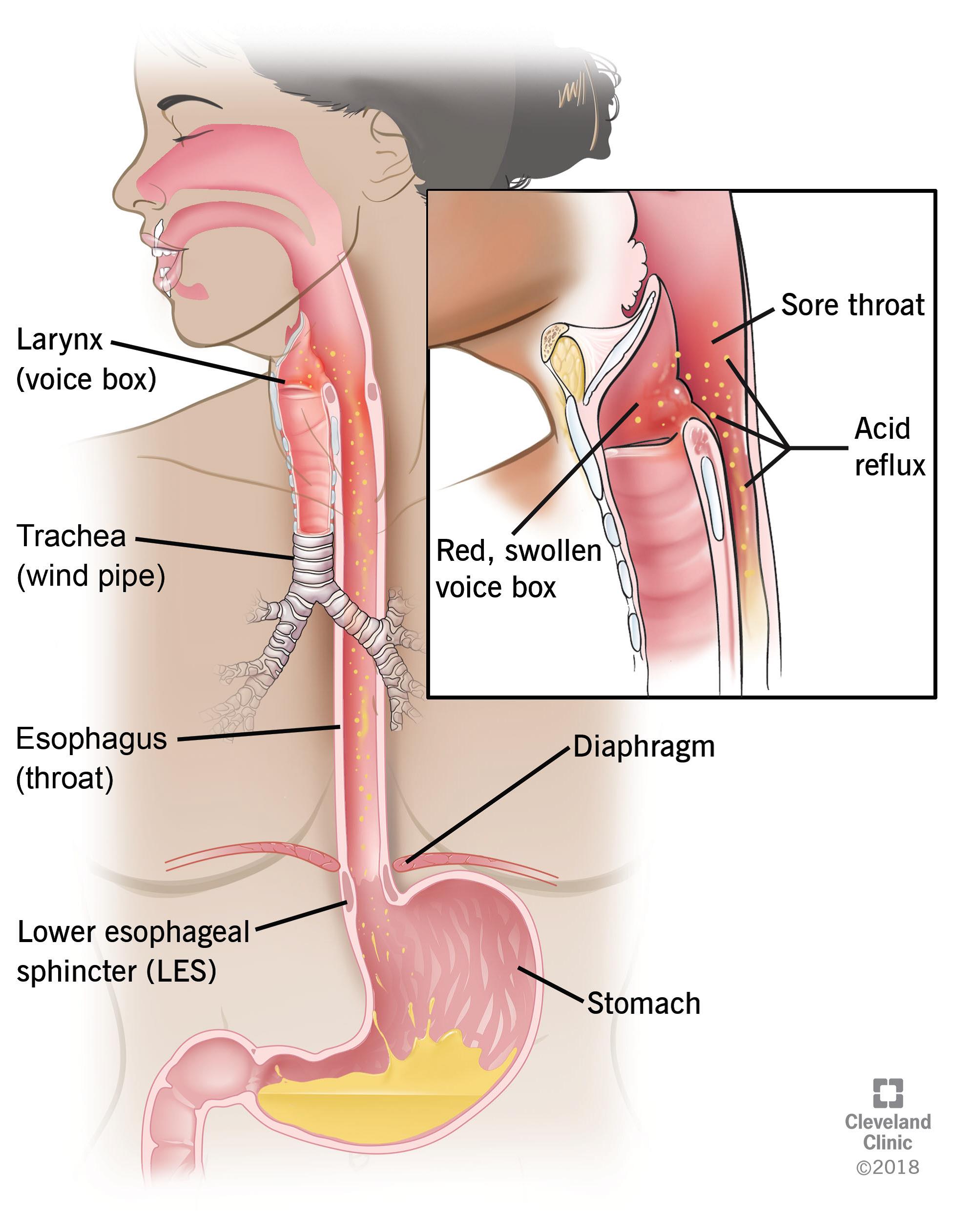
The esophagus is the hollow, muscular tube that moves food and liquid from the throat to the stomach. The wall of the esophagus is made up of several layers of tissue, including mucous membrane, muscle, and connective tissue. Esophageal cancer starts on the inside lining of the esophagus and spreads outward through the other layers as it grows.
ENLARGE
The esophagus and stomach are part of the upper gastrointestinal (digestive) system.
The two most common forms of esophageal cancer are named for the type of cells that become malignant (cancerous):
Squamous cell carcinoma: Cancer that forms in the thin, flat cells lining the inside of the esophagus. This cancer is most often found in the upper and middle part of the esophagus, but can occur anywhere along the esophagus. This is also called epidermoid carcinoma.
Adenocarcinoma: Cancer that begins in glandular cells. Glandular cells in the lining of the esophagus produce and release fluids such as mucus. Adenocarcinomas usually form in the lower part of the esophagus, near the stomach.
Signs and symptoms of esophageal cancer
These and other signs and symptoms may be caused by esophageal cancer or by other conditions. Check with your doctor if you have any of the following:
- Painful or difficult
- Swallowing.
- Weight loss.
- Pain behind
- The breastbone.
- Hoarseness and
- cough.
- Indigestion and heartburn.
A lump under the skin.
Tests that examine the esophagus
The following tests and procedures may be used:
Physical exam and health history: An exam of the body to check general signs of health, including checking for signs of disease, such as lumps or anything else that seems unusual. A history of the patient’s health habits and past illnesses and treatments will also be taken.
Chest x-ray: An x-ray of the organs and bones inside the chest. An x-ray is a type of energy beam that can go through the body and onto film, making a picture of areas inside the body.
Esophagoscopy: A procedure to look inside the esophagus to check for abnormal areas. An esophagoscope is inserted through the mouth or nose and down the throat into the esophagus. An esophagoscope is a thin, tube-like instrument with a light and a lens for viewing. It may also have a tool to remove tissue samples, which are checked under a microscope for signs of cancer. When the esophagus and stomach are looked at, it is called an upper endoscopy.
Biopsy: The removal of cells or tissues so they can be viewed under a microscope by a pathologist to check for signs of cancer. The biopsy is usually done during an esophagoscopy. Sometimes a biopsy shows changes in the esophagus that are not cancer but may lead to cancer.
Certain factors affect prognosis
The prognosis and treatment options depend on the following:
- The stage of the cancer (whether it affects part of the esophagus, involves the whole esophagus, or has spread to other places in the body).
Whether the tumor can be completely removed by surgery.
The patient’s general health. - When esophageal cancer is found very early, there is a better chance of recovery. Esophageal cancer is often in an advanced stage when it is diagnosed. At later stages, esophageal cancer can be treated but rarely can be cured. Taking part in one of the clinical trials being done to improve treatment should be considered. Information about ongoing clinical trials is available from the NCI website.
After esophageal cancer has been diagnosed
The process used to find out if cancer cells have spread within the esophagus or to other parts of the body is called staging. The information gathered from the staging process determines the stage of the disease. It is important to know the stage in order to plan treatment. The following tests and procedures may be used in the staging process:
Endoscopic ultrasound (EUS): A procedure in which an endoscope is inserted into the body, usually through the mouth or rectum. For esophageal cancer, the endoscope is inserted through the mouth. An endoscope is a thin, tube-like instrument with a light and a lens for viewing. A probe at the end of the endoscope is used to bounce high-energy sound waves (ultrasound) off internal tissues or organs and make echoes. The echoes form a picture of body tissues called a sonogram. A biopsy may also be done. This procedure is also called endosonography.
CT scan (CAT scan): A procedure that makes a series of detailed pictures of areas inside the body, such as the chest, abdomen, and pelvis, taken from different angles. The pictures are made by a computer linked to an x-ray machine. A dye may be injected into a vein or swallowed to help the organs or tissues show up more clearly. This procedure is also called computed tomography, computerized tomography, or computerized axial tomography.
PET scan (positron emission tomography scan): A procedure to find malignant tumor cells in the body. A small amount of radioactive glucose (sugar) is injected into a vein. The PET scanner rotates around the body and makes a picture of where glucose is being used in the body. Malignant tumor cells show up brighter in the picture because they are more active and take up more glucose than normal cells do. A PET scan and CT scan may be done at the same time. This is called a PET-CT.
MRI (magnetic resonance imaging): A procedure that uses a magnet, radio waves, and a computer to make a series of detailed pictures of areas inside the body. This procedure is also called nuclear magnetic resonance imaging (NMRI).
Thoracoscopy: A surgical procedure to look at the organs inside the chest to check for abnormal areas. An incision (cut) is made between two ribs and a thoracoscope is inserted into the chest. A thoracoscope is a thin, tube-like instrument with a light and a lens for viewing. It may also have a tool to remove tissue or lymph node samples, which are checked under a microscope for signs of cancer. In some cases, this procedure may be used to remove part of the esophagus or lung.
Laparoscopy: A surgical procedure to look at the organs inside the abdomen to check for signs of disease. Small incisions (cuts) are made in the wall of the abdomen and a laparoscope (a thin, lighted tube) is inserted into one of the incisions. Other instruments may be inserted through the same or other incisions to perform procedures such as removing organs or taking tissue samples to be checked under a microscope for signs of disease.
Ultrasound exam: A procedure in which high-energy sound waves (ultrasound) are bounced off internal tissues or organs, such as those in the neck, and make echoes. The echoes form a picture of body tissues called a sonogram. The picture can be printed to be looked at later.