History
Most patients with frictional keratosis are free of symptoms, with the exception of those with aggressive cheek and lip biting habits. In some individuals who repeatedly traumatize the tissues, tenderness, swelling, and a burning sensation may be presenting symptoms. Patients with persistent cheek and lip biting habits tend to have increased stress and psychologic disorders.
A patient may notice a thickening or roughness of the involved mucosal site, or frictional keratosis may be discovered as an incidental finding during a routine oral examination. Individuals with a cheek and lip biting habit often report they are able to remove thin strands or tags of mucosa from the involved site.
Patients may report that they are aware of sucking the mucosa or thrusting their tongue against their teeth. Some patients report that their cheeks and tongue feel swollen. Occasionally, the affected fungiform papillae in persons with a tongue biting or thrusting habit may be tender and sometimes associated with a burning sensation.
When the gingival tissues are involved, patients may report using a medium- or hard-bristled toothbrush or other oral hygiene aids. In some instances, patients give a history of wearing orthodontic appliances or removable full or partial dental prostheses that may traumatize the soft tissues. [20] Occasionally, ill-fitting or broken mouthguards or occlusal splints irritate the oral mucosa, resulting in frictional keratosis.
Sucking on the cheeks, lips, or sides of the tongue may be a habit to relieve the discomfort from temporomandibular disorder or burning mouth syndrome. Forceful or aberrant nutritional sucking on the nipple of the bottle or breast may result in calluses on the lips of infants.
In rare examples, individuals may give a history of picking the oral mucosa with long fingernails or some other external object.
Physical Examination
The first step in the identification of white patches suspected of being associated with physical trauma is to use a 2 X 2-inch sterile gauze to wipe off the lesion or lesions. If the patch is not easily wiped off, this suggests the presence of hyperkeratinization.
The lips, the lateral margins of the tongue, the buccal mucosa (mainly along the occlusal line), and the edentulous alveolar ridges are the most common sites to find frictional keratosis and its variants. Typically, the lesions appear as distinct, focal, and translucent-to-opaque white asymptomatic patches with sharply delineated borders. The surface of a lesion may appear irregular and feel rough to the tongue.
Slight variations in the clinical presentation are directly related to the nature and the source of the physical trauma.
One of the more common presentations of frictional keratosis is the linea alba (white line). This feature manifests as a horizontal thickening of the buccal mucosa along the occlusal line of the teeth. Linea alba is thought to result from chronic cheek biting or sucking of these tissues (see images below).
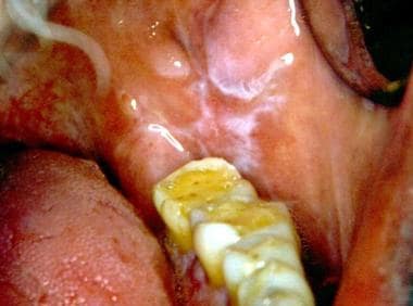 The white line observed on the cheek is level with the biting plane of the teeth. The wear on the occlusal surfaces of the molar teeth suggests that the patient had a habit of bruxism. Courtesy of Catherine M. Flaitz, DDS and Alfredo Aguirre, DDS.
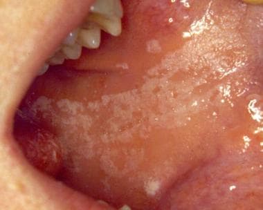 This wider area of roughened mucosa is typical of those produced by the habit of cheek biting or nibbling. Courtesy of Catherine M. Flaitz, DDS and Alfredo Aguirre, DDS.
In one patient, the surface of the last molar tooth showed considerable occlusal wear, which is evidence that the patient had the habit of grinding his teeth (see first image above). This habit most probably led to the biting of the cheek mucosa.
Occasionally, the line reflects the irregularity of the adjacent teeth and has a somewhat scalloped appearance (see image below).
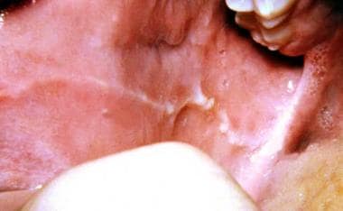 Prominent linea alba with evidence of cheek biting. The white line shows a slightly scalloped appearance, which correlates with the buccal surfaces of the teeth against which the mucosa is rubbed. Courtesy of Catherine M. Flaitz, DDS and Alfredo Aguirre, DDS.
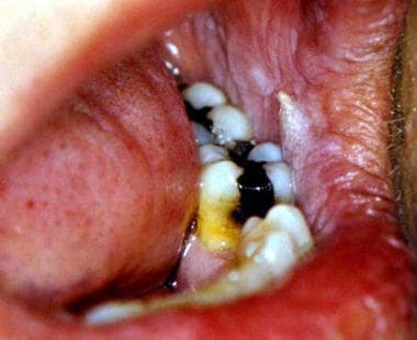
Occasionally, the frictional line is somewhat more diffuse, and this type of change is more likely to be associated with the habit of cheek chewing, also known as morsicatio buccarum (see images below), rather than the occasional accidental friction of teeth against the mucosa during the normal eating process. These white patches are associated with either a conscious or an unconscious chronic oral habit.
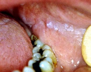 This frictional keratotic line shows a roughened surface. A thicker patch of mucosa is at the anterior end (under the tongue blade edge). This area is exactly level with the occlusal plane and was being chewed constantly by the patient. Courtesy of Catherine M. Flaitz, DDS and Alfredo Aguirre, DDS.
 Anterior rough surface area at the occlusal plane of the teeth. Courtesy of Catherine M. Flaitz, DDS and Alfredo Aguirre, DDS.
The effects of the habit of chronic biting may also manifest on the anterior and lateral borders of the tongue and appear as white, shaggy or mildly wrinkled plaques (see image below).
 Oral frictional hyperkeratosis of the lateral border of the tongue from chronic biting habit. Courtesy of Catherine M. Flaitz, DDS and Alfredo Aguirre, DDS.
A frictional keratosis lesion may be elevated from the surface, and patients may find that they develop the habit of nibbling further at these thickened mucosal sites. The first image below shows a frictional keratosis lesion that displays marked keratinization. The patient admitted to nibbling at the thickened mucosa (see second image below), which, in turn, made it thicker and easier to feel and, therefore, encouraged further nibbling.
This frictional keratotic line shows a roughened surface. A thicker patch of mucosa is at the anterior end (under the tongue blade edge). This area is exactly level with the occlusal plane and was being chewed constantly by the patient. Courtesy of Catherine M. Flaitz, DDS and Alfredo Aguirre, DDS.
Anterior rough surface area at the occlusal plane of the teeth. Courtesy of Catherine M. Flaitz, DDS and Alfredo Aguirre, DDS.
Occasionally, patchy erythema with or without petechiae is observed with recent trauma to the site.
Lesions associated with a tongue thrusting habit often demonstrate prominent crenations of the lateral tongue. In addition, the affected fungiform papillae may be red and enlarged from the chronic irritation.
-
The white line observed on the cheek is level with the biting plane of the teeth. The wear on the occlusal surfaces of the molar teeth suggests that the patient had a habit of bruxism. Courtesy of Catherine M. Flaitz, DDS and Alfredo Aguirre, DDS.
-
Prominent linea alba with evidence of cheek biting. The white line shows a slightly scalloped appearance, which correlates with the buccal surfaces of the teeth against which the mucosa is rubbed. Courtesy of Catherine M. Flaitz, DDS and Alfredo Aguirre, DDS.
-
This wider area of roughened mucosa is typical of those produced by the habit of cheek biting or nibbling. Courtesy of Catherine M. Flaitz, DDS and Alfredo Aguirre, DDS.
-
This frictional keratotic line shows a roughened surface. A thicker patch of mucosa is at the anterior end (under the tongue blade edge). This area is exactly level with the occlusal plane and was being chewed constantly by the patient. Courtesy of Catherine M. Flaitz, DDS and Alfredo Aguirre, DDS.
-
Anterior rough surface area at the occlusal plane of the teeth. Courtesy of Catherine M. Flaitz, DDS and Alfredo Aguirre, DDS.
-
Oral frictional hyperkeratosis of the lateral border of the tongue from chronic biting habit. Courtesy of Catherine M. Flaitz, DDS and Alfredo Aguirre, DDS.
-
Oral frictional hyperkeratosis of the attached maxillary gingiva from inappropriate toothbrushing technique. Courtesy of Catherine M. Flaitz, DDS and Alfredo Aguirre, DDS.
-
Oral frictional hyperkeratosis of the retromolar pad is also referred to as a ridge callus. This lesion is caused by masticatory irritation. Courtesy of Catherine M. Flaitz, DDS and Alfredo Aguirre, DDS.
-
Low-power view of stratified squamous epithelium with marked hyperkeratinization, acanthosis, and a prominent granular cell layer. Courtesy of Catherine M. Flaitz, DDS and Alfredo Aguirre, DDS.
-
High-power view of the surface keratin layer and a prominent granular cell layer. Courtesy of Catherine M. Flaitz, DDS and Alfredo Aguirre, DDS.


