History
The most common complaint is that of an asymptomatic or mildly tender, solitary, nonhealing ulcer of variable duration. The lesion may be present for as short as 1 week or for 12 months or longer. Patients with early ulcers often report pain and severe discomfort. Patients may have a history of trauma to the affected area. Depending on the location of the ulcer, other signs and symptoms may include dysphagia, odynophagia, dysphonia, and dyspnea. Occasionally, patients may present with a history of recent weight loss.
Infants with Riga-Fede disease often experience discomfort while breastfeeding, and they may fail to thrive in the postnatal period.
Physical Examination
Clinical appearance
Eosinophilic ulcer typically presents as an irregular, solitary ulcer with a fibrinous membrane on the surface. A zone of erythema surrounds the solitary ulcer. The margins of the lesion are often raised and usually indurated. [10] Purulence may be noted emanating from the ulcer.
Eosinophilic ulcers may be a few millimeters to as large as 7-8 cm in greatest dimension.
In rare reports, multiple synchronous or metachronous lesions have been identified.
Occasional ulcers may be macular, whereas others may present as nonspecific erythroplakic or leukoplakic lesions.
In rare cases, an eosinophilic ulcer may present as an elevated, smooth mass that is free of ulceration; however, biopsy reveals the underlying, characteristic, invasive cellular proliferation. In some of these cases, the overlying epithelium may have regenerated without resolution of the underlying inflammation. [4]
Mucosal sites
Any mucosal surface can be affected; however, the tongue is the most common location, accounting for 60% of reported cases. [19, 20] The lateral and dorsal surfaces are usually affected because these are the areas most often traumatized.
Lesions on the ventral surface of the tongue more commonly are observed in infants because of contact with the adjacent mandibular incisors during breastfeeding.
The dorsal surface of the tongue may also be affected in infants because of irritation associated with maxillary incisors. The buccal mucosa and mucobuccal fold are also particularly susceptible to ulceration; lesions in these locations account for 24% of reported cases.
Eosinophilic ulcers have also been reported (in decreasing order of frequency) on the lips, gingiva, palate, floor of the mouth, and retromolar area.
In extremely rare cases, cervical lymphadenopathy is reported.
(See the images below.)
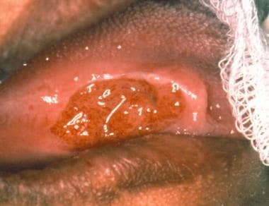 A 47-year-old African American woman with an eosinophilic ulcer on the lateral surface of the tongue. The anterior border of the lesion is raised. Courtesy of Dr Paul D. Freedman.
 Raised, indurated, nonhealing ulcer on the lateral surface of the tongue. The lesion was related to an adjacent fractured tooth. Courtesy of Dr Paul D. Freedman.
 Ulcer on the ventrolateral surface of the tongue. The differential diagnosis should include squamous cell carcinoma or an infectious etiology. Courtesy of Dr Paul D. Freedman.

Causes
Common causes of oral trauma include the following:
-
Self-inflicted injury in which the patient accidentally or deliberately traumatizes the mucosa
-
Injury due to sharp-edged teeth or food
-
Injury due to neonatal or natal teeth (Riga-Fede disease)
-
Toothbrush abrasion
-
Injury due to ill-fitting dentures
-
Injury due to orthodontic or occlusal appliances
-
Iatrogenic injuries (eg, those that occur during dental procedures, such as anesthetic necrosis that occurs during intubation for surgery)
-
Injuries due to accidents
Certain patients may be inherently predisposed to the development of eosinophilic ulcers, although this factor remains controversial.
The role of drug reactions, if any, is unclear.
Medical conditions or therapeutic regimens that predispose an individual to immune suppression may also delay healing.
-
A 47-year-old African American woman with an eosinophilic ulcer on the lateral surface of the tongue. The anterior border of the lesion is raised. Courtesy of Dr Paul D. Freedman.
-
Raised, indurated, nonhealing ulcer on the lateral surface of the tongue. The lesion was related to an adjacent fractured tooth. Courtesy of Dr Paul D. Freedman.
-
Ulcer on the ventrolateral surface of the tongue. The differential diagnosis should include squamous cell carcinoma or an infectious etiology. Courtesy of Dr Paul D. Freedman.
-
Lesion on the lateral surface of the tongue. Courtesy of Dr Paul D. Freedman.
-
Low-power view showing an ulcerated surface epithelium with a dense cellular inflammatory infiltrate underlying the mucosal surface (original magnification X40). Courtesy of Dr Paul D. Freedman.
-
Cellular infiltrate composed mainly of large mononuclear cells, including histiocytes and submucosal dendrocytes, eosinophils, and scattered T lymphocytes (original magnification X400). Courtesy of Dr Paul D. Freedman.
-
Inflammatory infiltrate extending through and between muscle bundles (original magnification X400). Courtesy of Dr Paul D. Freedman.


