History
Patient history can reliably suggest the presence of scabies. Lesion distribution and intractable pruritus that is worse at night, as well as scabies symptoms in close contacts (including multiple family members), should immediately rank scabies at the top of the clinical differential diagnosis.
Lesion distribution differs in adults and children. Adults manifest lesions primarily on the following parts of the body (see the images below):
-
Flexor aspects of the wrists
-
Interdigital web spaces of the hands
-
Dorsal feet
-
Axillae
-
Elbows
-
Waist
-
Buttocks
-
Genitalia
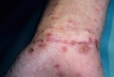 Erythematous papules and papulovesicles on the flexor wrist. Courtesy of Kenneth E. Greer, MD.


Pruritic papules and vesicles on the scrotum and penis in men and areolae in women are highly characteristic.
Infants and young children may develop lesions diffusely, but unlike in adults, lesions are common on the face, scalp, neck, palms, and soles. [34]
All cutaneous sites are susceptible in immunocompromised and elderly patients, who often have a history of a widespread, pruritic, eczematous eruption. The same is true in patients with existing dermatologic, particularly atopic, diseases, in whom scabies may exacerbate or complicate these conditions. [34]
Patients who have had scabies misdiagnosed may have been treated with topical corticosteroids. This delays the correct diagnosis as the lesions may take on the incognito form in which lesions might appear as vesicles, pustules, or nodules and whose distribution could become diffuse. [35, 36]
Signs and symptoms of pruritus tend to crescendo progressively over 2-3 weeks before compelling the patient to seek medical attention. (However, debilitated or immunocompromised patients may not have the urge to scratch. [37] )
Scabies appears to occur in clusters. If there is an outbreak in the community, consider scabies in an individual presenting with rash and itching.
Physical Examination
Clinical findings include primary and secondary lesions. Primary lesions are the first manifestation of the infestation and typically include small papules, vesicles, and burrows. Secondary lesions are the result of rubbing and scratching, and they may be the only clinical manifestation of the disease. If so, the diagnosis must be inferred by the history, lesion distribution, and accompanying symptoms. [38]
Primary scabies lesions
Burrows are a pathognomonic sign and represent the intraepidermal tunnel created by the moving female mite. They appear as serpiginous, grayish, threadlike elevations in the superficial epidermis, ranging from 2-10 mm long, as seen in the image below.

They are not readily apparent and must be actively sought. A black dot may be seen at one end of the burrow, indicating the presence of a mite. High-yield locations for burrows include the following:
-
Webbed spaces of the fingers
-
Flexor surfaces of the wrists
-
Elbows
-
Axillae
-
Belt line
-
Feet
-
Scrotum (men)
-
Areolae (women)
Classic scabies typically has a distribution involving the axillae, elbow flexures, wrists and hands, and genital area. This is commonly known as the circle of Hebra.
In geriatric patients, scabies demonstrates a propensity for the back, often appearing as excoriations. In infants and small children, burrows are commonly located on the palms and soles, as in the image below. In immunocompromised patients, bullous lesions may be observed.
 A subtle linear burrow accompanied by erythematous papules on the sole of the foot in a child with scabies. Courtesy of Kenneth E. Greer, MD.
One- to 3-mm erythematous papules and vesicles are seen in typical distributions in adults. The vesicles are discrete lesions filled with clear fluid, although the fluid may appear cloudy if the vesicle is more than a few days old, as in the image below.
 Papulovesicles and nodules on the palm in a patient with scabies. Courtesy of Kenneth E. Greer, MD.
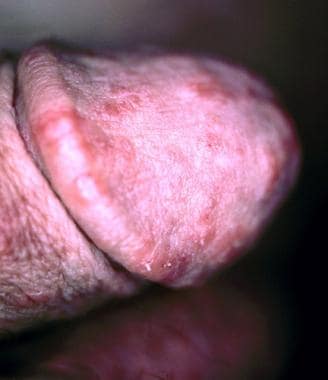
Papules rarely contain mites and most likely represent a hypersensitivity reaction. Papules are common on the shaft of the penis in men and on the areolae in women. (See the images below.)

 Scabietic papules on the penile shaft and scrotum. Courtesy of Kenneth E. Greer, MD.
Unlike adults, children commonly present with facial and neck involvement. In very young children and infants, a widespread, eczematous eruption primarily on the trunk is common, as in the image below. In addition, infants may have 1- to 3-mm papules, vesicles, and pustules on the palms and soles.
 Widespread eruption on the back of an infant with scabies. Courtesy of Kenneth E. Greer, MD.
Nodular scabies
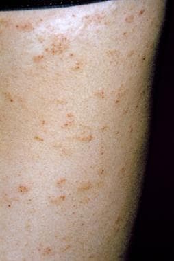
Nodules occur in 7-10% of patients with scabies, particularly young children. In neonates unable to scratch, pinkish brown nodules ranging in size from 2-20 mm in diameter may develop, as demonstrated in the images below. Mites are rarely found within the nodules.


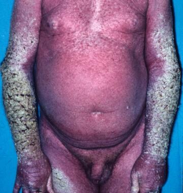
Crusted scabies
Crusted scabies, previously referred to as Norwegian scabies, manifests with marked thickening and crusting of the skin. Lesions are often hyperkeratotic and crusted and cover large areas. Marked scaling is common, and pruritus may be minimal or absent. Nail dystrophy (subungual hyperkeratosis) and scalp lesions may be present.
The hands and arms are the usual locations for lesions, but all sites are vulnerable. Mites can number in the thousands to millions in this form of scabies. (See the images below.)
Secondary scabies lesions
These lesions result from scratching, secondary infection, and/or the host’s immune response against the scabies mites and their products. Characteristic findings include the following [1, 2, 3] :
-
Excoriations (see the image below)
-
Widespread eczema
-
Honey-colored crusting
-
Postinflammatory hyperpigmentation
-
Erythroderma
-
Prurigo nodules
-
Frank pyoderma
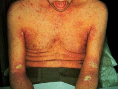
 Scabies. Erythematous vesicles and papules are present on torso extremities, some with adjacent linear excoriations.
-
Scabies mite scraped from a burrow (original magnification, 400X).
-
A typical linear burrow on the flexor forearm. Courtesy of Kenneth E. Greer, MD.
-
A subtle linear burrow accompanied by erythematous papules on the sole of the foot in a child with scabies. Courtesy of Kenneth E. Greer, MD.
-
Erythematous papules and papulovesicles on the flexor wrist. Courtesy of Kenneth E. Greer, MD.
-
Scabies on the penile shaft and glans. Courtesy of William D. James, MD.
-
Scabietic papules on the penile shaft and scrotum. Courtesy of Kenneth E. Greer, MD.
-
Widespread eruption on the back of an infant with scabies. Courtesy of Kenneth E. Greer, MD.
-
Nodular scabies in an infant. Courtesy of Kenneth E. Greer, MD.
-
Nodular scabies. Courtesy of Kenneth E. Greer, MD.
-
Crusted scabies. Courtesy of William D. James, MD.
-
Crusted scabies. Courtesy of Kenneth E. Greer, MD.
-
Scabies preparation demonstrating a mite and ova. Courtesy of William D. James, MD.
-
Scabies. Erythematous vesicles and papules are present on torso extremities, some with adjacent linear excoriations.
-
In routine scabies, a single mite is seen. Eosinophilic spongiosis may be present (hematoxylin and eosin; original magnification, 400X).
-
Scabies mite in the stratum corneum. Courtesy of William D. James, MD.
-
In crusted scabies, sections show multiple mites (arrows) within the hyperkeratotic stratum corneum. The epidermis is spongiotic (hematoxylin and eosin; original magnification, 100X).
-
Scabies. Courtesy of William D. James, MD.
-
Scabies in the interdigital web spaces. Courtesy of William D. James, MD.
-
Papulovesicles and nodules on the palm in a patient with scabies. Courtesy of Kenneth E. Greer, MD.
-
Scabies on buttocks. Courtesy of William D. James, MD.
-
Scabies on penis. Courtesy of Hon Pak, MD.


