History
Patients report pruritus of the hands and feet with a sudden onset of vesicles. Burning pain or pruritus occasionally may be experienced before vesicles appear. Tiny vesicles erupt first along lateral aspects of the fingers and then on the palms or soles. Palms and soles may be red and wet with perspiration. The vesicles usually persist for 3-4 weeks. Vesicle outbreaks may occur in waves. A photo-induced form of hand dermatitis resembling dyshidrotic eczema has been described. [15]
Dyshidrotic eczema episodes vary in frequency from once per month to once per year. Patients with dyshidrotic eczema may report a variety of factors that possibly are related to eruptions, as follows:
-
Emotional stress
-
Personal or familial atopic diathesis (eg, asthma, hay fever, sinusitis)
-
Certain work exposures (eg, cobalt) and/or recreational exposures
-
Recent exposure to contact allergens (eg, nickel, balsams, paraphenylenediamine, chromate, sesquiterpene lactones) before condition flares
-
Exposure to contact irritants before condition flares
-
Recent exposure to costume jewelry (patients with palmar pompholyx and allergy to nickel)
-
Human immunodeficiency virus (HIV) infection
Two cases were reported of HIV-positive patients who developed dyshidrotic eczema as an immune reconstitution inflammatory syndrome shortly after highly active antiretroviral therapy. [22] Pompholyx has also been described as a manifestation of symptomatic HIV infection, including in individuals who do not respond to topical and systemic therapies and whose condition resolves only after the initiation of combination antiretroviral therapy. [23]
One publication provides an algorithm for the diagnosis of chronic hand dermatitis, which offers an easy way of classifying these skin conditions, helping in the decision making when choosing treatment modalities. [24]
Physical Examination
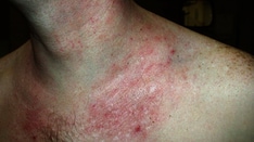
Symmetrical crops of clear vesicles and/or bullae (blisters) on the palms and lateral aspects of the fingers characterize dyshidrotic eczema. The feet, the soles, and the lateral aspects of toes also may be affected. (See the images below.)
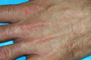 Dyshidrotic eczema (pompholyx). Small, discrete, coalesced vesicles on the dorsal hand.
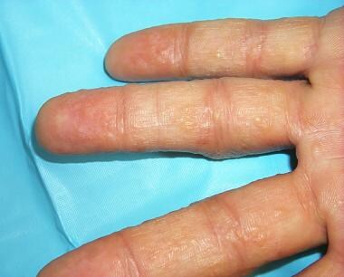
 Dyshidrotic eczema (pompholyx). Small, discrete, coalesced vesicles on the fingers.
In mildly affected patients, vesicles are present only on the lateral aspects of the fingers and, occasionally, involve feet and toes. (See the image below.)

Vesicles are deep seated and have a tapiocalike appearance, without surrounding erythema. They may become large, form bullae, and become confluent. Vesicles typically resolve without rupturing, followed by desquamation. (See the image below.)
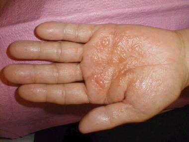 Dyshidrotic eczema (pompholyx). Close-up view of tense vesicles and bullae of the palm. Courtesy of Norman Minars, MD, University of Miami, Department of Dermatology & Cutaneous Surgery.
In 80% of patients, only the hands are involved, while in 10% of patients, the disease affects only the feet, and in another 10% of patients, the hands and feet are involved together. (See the images below.)
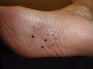 Dyshidrotic eczema (pompholyx). Discrete yellow pustules on the sole of the foot. Courtesy of Norman Minars, MD, University of Miami, Department of Dermatology & Cutaneous Surgery.
 Dyshidrotic eczema (pompholyx). Palms and soles of a patient with a dyshidrosis flare. The patient unroofed a large bulla on the right sole.
With long-standing disease, patients' fingernails may reveal dystrophic changes (eg, irregular, transverse ridging; pitting; thickening; discoloration). Interdigital maceration and desquamation of the interdigital spaces often are present, despite the possible absence of a dermatophyte infection. Vesicles and/or bullae may become infected secondarily, and pustular lesions may be present. Cellulitis and lymphangitis may develop.
Severity index
The Dyshidrotic Eczema Area and Severity Index was developed based on severity grades for the number of vesicles per square centimeter, erythema, desquamation, itch, and the extent of affected areas. [25] The index was found to be a simple standardized method for assessing the condition and was used to assess disease severity and treatment effectiveness in two clinical studies. Further evaluation with larger patient groups is needed.
Complications
Secondary bacterial infection of dyshidrotic eczema vesicles or bullae can result in cellulitis, lymphangitis, and septicemia (rare). Dystrophic nail changes may develop, with the occurrence of transverse ridging, thickening, discoloration, and pitting. Dyshidrotic eczema has no associated mortality, although some severe cases can become debilitating. A 2015 study determined that dyshidrosis is a risk factor in the development of herpes zoster. [26]
-
Dyshidrotic eczema (pompholyx). Tense vesicles and bullae on the palm. Courtesy of Norman Minars, MD, University of Miami, Department of Dermatology & Cutaneous Surgery.
-
Dyshidrotic eczema (pompholyx). Close-up view of tense vesicles and bullae of the palm. Courtesy of Norman Minars, MD, University of Miami, Department of Dermatology & Cutaneous Surgery.
-
Dyshidrotic eczema (pompholyx). Discrete yellow pustules on the sole of the foot. Courtesy of Norman Minars, MD, University of Miami, Department of Dermatology & Cutaneous Surgery.
-
Dyshidrotic eczema (pompholyx). Multiple tense vesicles on the palm.
-
Dyshidrotic eczema (pompholyx). Small tense vesicles on the fingers.
-
Dyshidrotic eczema (pompholyx). Small, discrete, coalesced vesicles on the dorsal hand.
-
Dyshidrotic eczema (pompholyx). Small, discrete, coalesced vesicles on the fingers.
-
Dyshidrotic eczema (pompholyx). Palms and soles of a patient with a dyshidrosis flare. The patient unroofed a large bulla on the right sole.
-
Dyshidrotic eczema (pompholyx). Small discrete vesicles of the lateral fingers.