Background
Toe amputation is a common procedure performed by a wide variety of healthcare providers. The vast majority of toe amputations are performed on patients with a diabetic foot. [1] Although regional variation is noted, most of these procedures are done by general, vascular, and orthopedic surgeons (particularly those subspecializing in foot and ankle surgery); in some countries, podiatrists are involved.
There are three broad indications for amputation of any body part, [2] as follows (see Indications):
-
Dead
-
Deadly
-
Dead loss
Before any amputation, the clinician should ensure that the patient’s medical circumstances have been optimized (ie, should "reverse the reversible"). With impending toe amputation, this step encompasses such measures as glycemic control and consideration of revascularization when severe macrovascular disease is contributing to ischemia.
The method of toe amputation (disarticulation vs osteotomy) and the level of amputation (partial or whole phalanx vs whole digit vs ray) depend on numerous circumstances but are mainly determined by the extent of disease and the anatomy. With any amputation, the degree of postoperative functional loss is generally proportional to the amount of tissue taken. The great toe is considered the most important of the toes in functional terms. Nevertheless, great-toe amputation can be performed with little resulting functional deficit.
Indications
A “dead” toe is one in which the blood supply is so completely impeded that infarction and necrosis develop (see the images below). Infarction results in dry gangrene, with nonviable tissue becoming dry and black in color (because of the presence of iron sulfide, a product of the hemoglobin released by lysed erythrocytes).
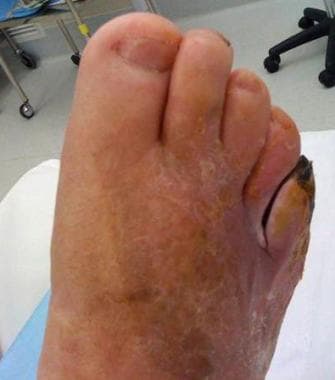
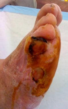
In Western societies, a dead toe is most commonly seen as a complication of diabetes mellitus [3] and is due to a combination of macrovascular and microvascular disease. Other major risk factors for peripheral vascular disease (as for all atherosclerotic diseases) include smoking, hypertension, and hyperlipidemia. Rarely, pathologic processes confined to the microvasculature are to blame (eg, thromboangiitis obliterans [Buerger disease], vasospastic diseases, severe frostbite).
The “deadly” category generally refers to a more proximal disease processe that can result in systemic sequelae. For example, a large limb segment affected by wet gangrene (with macroscopic putrefaction and surrounding cellulitis) can be deadly if not addressed urgently. Malignancy may also necessitate amputation, though infrequently.
A toe is a “dead loss” when it is diseased to the point where it is irreparable (as with chronic osteomyelitis; in patients with mild or limited disease without sepsis, chronic osteomyelitis may be treatable by nonoperative means [4] ), it ceases to be functional (as with significant trauma), or it is impeding function (as with neuropathic pain).
Ray amputation has been performed to treat foot macrodactyly in children. [5]
Contraindications
The main contraindication for toe amputation is poor demarcation of infarcted tissue (patchy gangrene). If the borders of the infarcted area are unclear, the surgeon cannot know the true extent of the disease and hence cannot be certain of amputating to a region of adequate blood supply.
More broadly, amputation of any body part is contraindicated when it will result in a reduced quality of life in the setting of a limited life expectancy. However, this contraindication is not usually relevant to toe amputation.
Technical Considerations
Anatomy
The phalanges of the foot correspond, in number and general arrangement, with those of the hand; two are found in the great toe, and three are found in each of the other toes. However, the phalanges of the foot differ from the phalanges of the hand in terms of size, with the bodies of the phalanges of the foot being much reduced in length and, especially in the first row, laterally compressed.
The body of each proximal phalanx is similar to the metatarsals in being convex above and concave below. The base is concave for articulation with the respective metatarsal, and the head presents a trochlear surface for articulation with the second phalanx.
For more information about the relevant anatomy, see Foot Bone Anatomy.
Outcomes
Vassallo et al performed a single-center study (N = 81) aimed at determining healing, reulceration, reamputation, and mortality rates at 1 year after toe amputations in patients with type 2 diabetes mellitus. [6] In 12.4% of participants, the amputation site remained incompletely healed; in 80.2%, the amputation site was completely healed at 12 months. Only 20.9% had no complications in 12 months. Over the study period, 45.7% of subjects had had a new ulcer at a different site. Forty-eight participants (59.3%) underwent further surgery, either to revise the original amputation site (n = 31) or to amputate at a new site (n = 17). Mortality was 7.4%.
In a study of patients (N = 146; mean age, 65 y) who underwent amputation of the hallux (n = 55) or another toe (n = 91) for irreversible foot sepsis, Collins et al found that 43.2% required further major or minor ipsilateral digital amputation after the index toe amputation. [7] Rates of limb preservation were high, and most of the patients were alive at 5 years. No significant difference in outcome was noted between patients who underwent hallux amputation and those who underwent amputation of other toes. The only independent predictor of ipsilateral major amputation–free survival and overall survival in this study was increasing age.
A study by Speer et al (N = 116) found that diabetic patients were significantly more likely to require reoperations and reamputations after toe amputations than nondiabetic patients were. [8]
A retrospective study by Rolle et al (N = 375) examined risk factors for conversion of minor lower-extremity amputations to repeat minor amputations or major amputations. [9] Patients were divided into two groups: toe/ray amputations (n = 245) and midfoot amputations (n = 130). The toe/ray group was more likely to have a repeat minor amputation within 1 year and had a higher rate of wound healing at 90 days; the midfoot group was more likely to undergo a major lower-extremity amputation within 1 year. There was no significant difference in mortality between the two groups.
A retrospective study by Víquez-Molina et al (N = 175) addressed risk factors associated with the failure of one- to three-toe amputation in patients with diabetic foot infection. [10] Failed amputation, necessitating a more proximal amputation, occurred in 53 patients (30.3%). Factors associated with a higher rate of amputation failure were severe infection, isolation of Pseudomonas aeruginosa and Escherichia coli, and prolonged prothrombin time; factors associated with a lower rate were obesity and elevated hemoglobin level.
-
Markup for second-toe amputation (fish-mouth skin excision): anterior view of (dry) gangrenous toe.
-
Markup for second-toe amputation: posterior view.
-
Toe excised. Note shiny cartilage cap on metatarsal head.
-
Metatarsal head cartilage removed.
-
Dressing suggestion: wound packed with calcium-alginate dressing, paraffin gauze on top (crepe bandage to finish).
-
Adjacent toe amputations: anterior view of markup.
-
Fourth metatarsal head cartilage. (For removal, see video.)
-
Gangrenous fifth toe. Dubious perfusion in fourth toe.
-
Lateral two toes for amputation (lateral view).
-
Lateral two toes amputated. Note alternative technique used here. Single larger wound is made rather than two smaller fish-mouth wounds; this is because skin bridge between toes was unlikely to have adequate perfusion.
-
Amputation of adjacent toes at metatarsophalangeal joint using fish-mouth incisions.
-
Revision of transmetatarsal amputation.