Life continues from one generation to the other as a result of replication, transcription, and translation of genetic code stored in DNA and RNA.
Genetic code contains the sequence of nucleotide bases within DNA and RNA. DNA consists of four nucleotide bases: adenine (A), guanine (G), cytosine (C), and thymine (T). The genetic code within DNA is transcribed to RNA which acts as a template for protein synthesis. RNA consists of the bases adenine, guanine, cytosine, and uracil (U). They code for amino acids that link together to form proteins. So genetic code acts as a set of instructions that assemble amino acids in the correct order to form proteins.
The genetic code is read in sets of three nucleotides and this sequence of three DNA or RNA nucleotides is known as codons. Each codon specifies a particular amino acid or acts as a signal to start or stop protein synthesis.
The first codon was discovered by Marshall Nirenberg and J. Heinrich Matthaei’s poly-U experiment which identified that a chain of three uracil units (UUU) in RNA acted as a code word for the amino acid phenylalanine. Nirenberg’s subsequent work, alongside a team of researchers, led to the identification of all 64 possible codons and their corresponding amino acids. Nirenberg was awarded the Nobel Prize in Physiology or Medicine in 1968, sharing the honor with Robert W. Holley and Har Gobind Khorana for their combined efforts in deciphering the genetic code.
Interesting Science Videos
What is a Codon Chart?
A codon chart or table is used as a reference tool that correlates specific codons with the corresponding amino acids they encode.
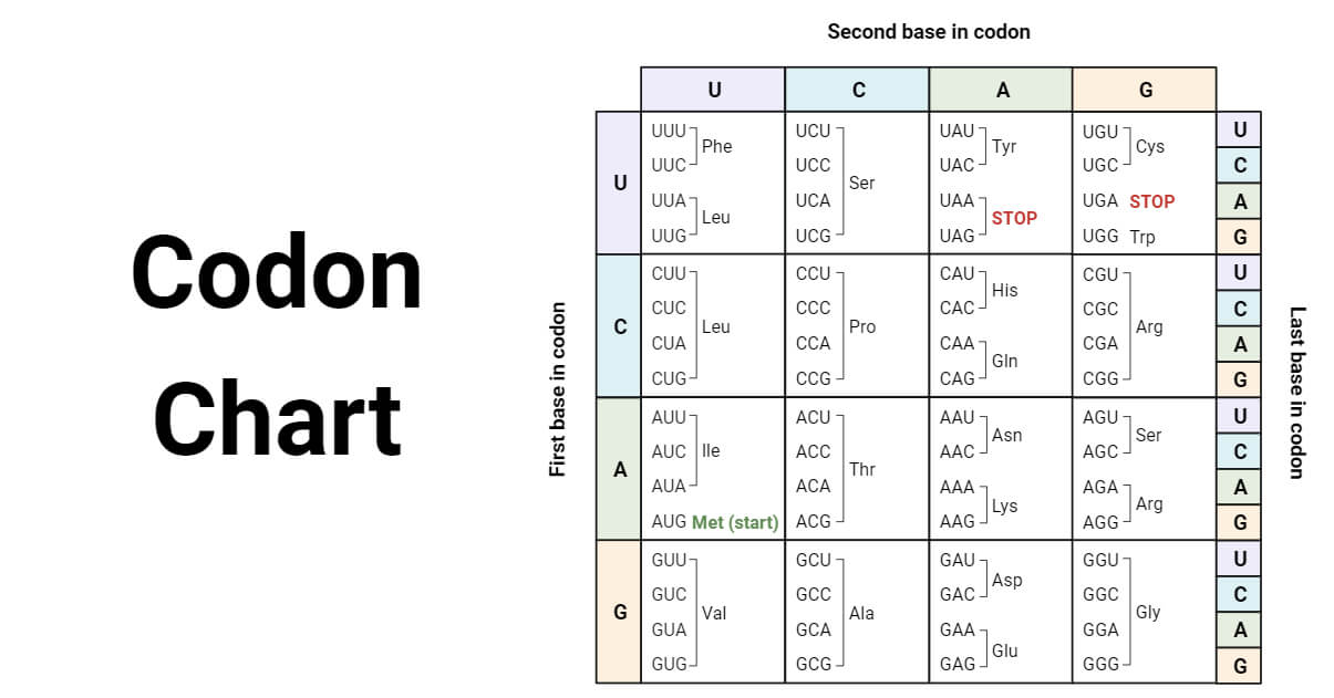
- The chart helps to decipher the genetic code and understand which amino acids are synthesized based on the sequence of nucleotides.
- In total, there are 64 possible codons. Out of these, 61 codons correspond to the 20 different amino acids and the remaining three are stop codons that signal the end of protein synthesis.
- In the chart, we can see that a single amino acid can be coded by multiple codons, so the genetic code is redundant or degenerate. For example, the codons CCU, CCC, CCA, and CCG all code for the same amino acid proline.
- The codon chart outlines the various codon combinations and their corresponding amino acids.
- To use the codon chart, for example, if the first codon position contains uracil (U), the second contains adenine (A), and the third contains cytosine (C), the resulting codon, UAC, represents the amino acid tyrosine.
Here is the list of the 20 amino acids, along with their three-letter and one-letter abbreviation:
| Amino Acid | Abbreviation | One-Letter Abbreviation |
| Alanine | Ala | A |
| Arginine | Arg | R |
| Asparagine | Asn | N |
| Aspartic acid | Asp | D |
| Cysteine | Cys | C |
| Glutamic acid | Glu | E |
| Glutamine | Gln | Q |
| Glycine | Gly | G |
| Histidine | His | H |
| Isoleucine | Ile | I |
| Leucine | Leu | L |
| Lysine | Lys | K |
| Methionine | Met | M |
| Phenylalanine | Phe | F |
| Proline | Pro | P |
| Serine | Ser | S |
| Threonine | Thr | T |
| Tryptophan | Trp | W |
| Tyrosine | Tyr | Y |
| Valine | Val | V |
Start Codons
- Start codons are codons within an mRNA molecule that indicate the start of protein translation.
- AUG is the most common start codon and serves as the start signal for translation initiation.
- In eukaryotes, AUG codes for the amino acid methionine (Met), while in prokaryotes, AUG codes for formyl methionine (fMet).
- Initiation factors and tRNA are involved in recognizing the start codon AUG and initiating the translation process.
- Although AUG is the primary start codon, alternative codons can also serve as start sites. These non-AUG start codons are relatively rare in eukaryotes.
- Prokaryotes like E. coli use various codons including AUG, GUG, and UUG as start codons.
Stop Codons
- Out of the total 64 codons, there are three codons that do not specify or correspond to any amino acids. These codons are known as stop codons.
- Stop codons, also called termination or nonsense codons, serve as signals to terminate the process of protein synthesis during translation.
- The three stop codons are UAA, UAG, and UGA.
- Stop codons were first discovered in 1965 by Sydney Brenner’s experiment on T4 bacteriophage.
- UAG was the first stop codon to be identified. It is also called the amber codon. UGA is called the opal codon, and UAA is called the ochre codon.
Codon Translation
- Codon translation is a crucial step in protein synthesis. Proteins are essential molecules in living organisms that are synthesized during translation.
- The genetic information stored in DNA is not directly used to build proteins; instead, it is first transcribed into RNA.
- DNA transcription is a critical step in protein synthesis where genetic instructions are copied from DNA to RNA. The enzyme RNA polymerase transcribes DNA into a single-stranded RNA molecule known as messenger RNA (mRNA).
- Protein synthesis occurs in the cytoplasm, where the mRNA, along with ribosomes and transfer RNA (tRNA), translates the transcribed genetic code into chains of amino acids.
- Translation involves reading each RNA codon and adding the corresponding amino acid to the chain of amino acids carried by tRNA molecules. This process stops when a stop codon is encountered in the mRNA sequence.
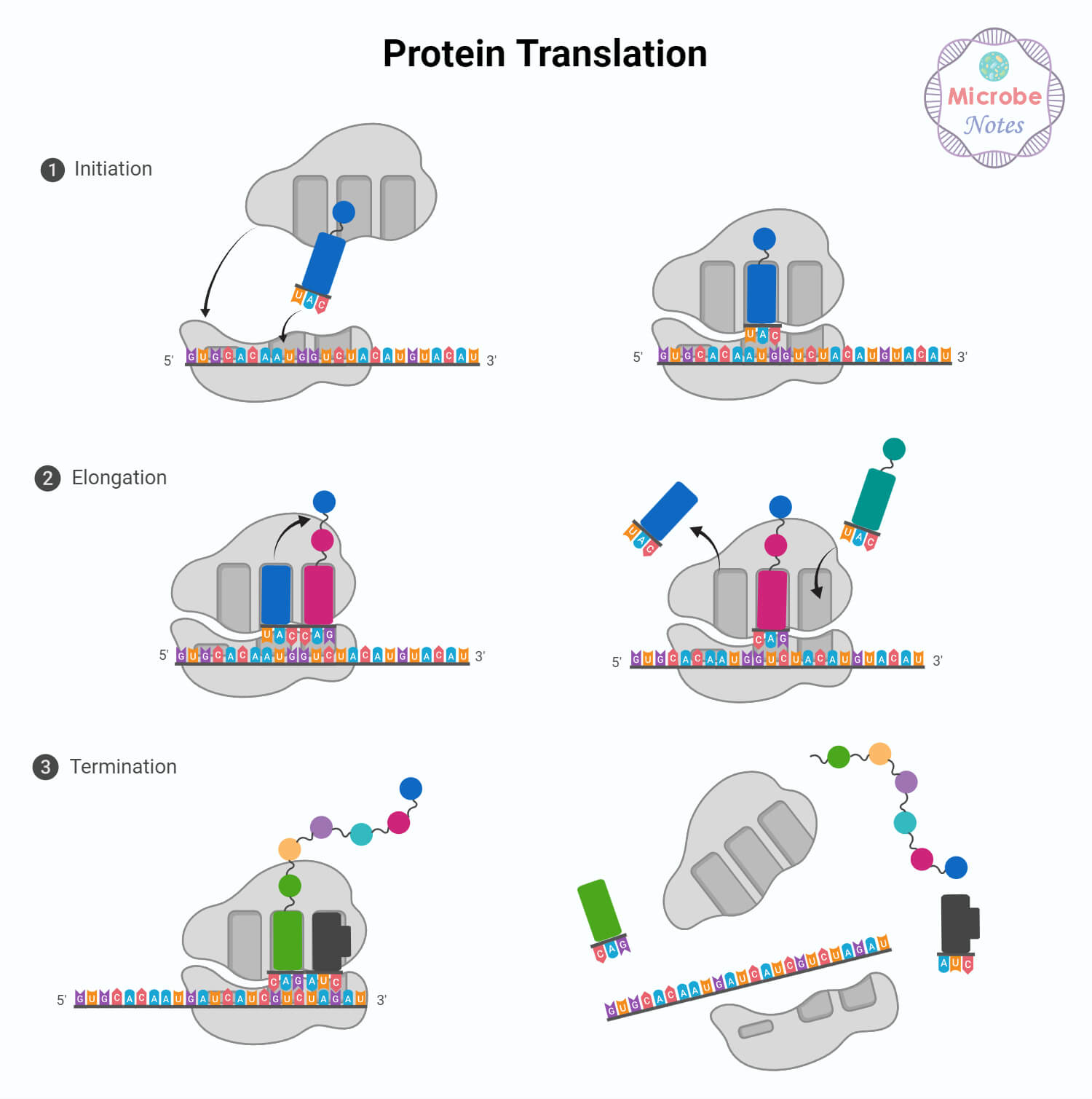
- The unique structure of tRNA helps in the precise matching between mRNA codon and the corresponding amino acid.
- The tRNA molecule, which resembles a cloverleaf shape, comprises four double-helical stems and three single-stranded loops. The central loop is referred to as the anticodon loop, as it carries a set of three nucleotides known as the anticodon.
- The tRNA anticodon sequence is complementary to the mRNA codon and this complementarity allows for a specific and accurate interaction between the anticodon and the codon.
- Once transcription and translation are complete, the newly formed chain of amino acids undergoes modifications to transform into a functional protein.
References
- American Chemical Society National Historic Chemical Landmarks. Deciphering the Genetic Code. http://www.acs.org/content/acs/en/education/whatischemistry/landmarks/geneticcode.html
- Bailey, Regina. “Understanding the Genetic Code.” ThoughtCo, Aug. 29, 2020, thoughtco.com/genetic-code-373449.
- Clancy, S. & Brown, W. (2008) Translation: DNA to mRNA to Protein. Nature Education 1(1):101
- Griffiths, A. J. F., Miller, J. H., Suzuki, D. T., Lewontin, R. C., Gelbart, W. M. (2000). An introduction to genetic analysis (7th ed.). New York: W. H. Freeman.
- https://www.genome.gov/genetics-glossary/Codon
- https://www.nature.com/scitable/definition/codon-155/
- https://www.nature.com/scitable/definition/genetic-code-13/
- Koonin, E. V., & Novozhilov, A. S. (2009). Origin and evolution of the genetic code: The universal enigma. Iubmb Life, 61(2), 99. https://doi.org/10.1002/iub.146
- Smith, A. (2008) Nucleic acids to amino acids: DNA specifies protein. Nature Education 1(1):126.
- The Information in DNA Determines Cellular Function via Translation | Learn Science at Scitable (nature.com)
- Translating the Code of Life and the Nobel Prize, 1962-1968 | Marshall W. Nirenberg – Profiles in Science (nih.gov)