CASE 10
Clinical Presentation
Antenatal US diagnosis of a brain anomaly included absent septum pellucidum and microcephaly. Early neonatal MRI study was performed for better malformation depiction and identification.
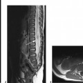
Figure 10A
Radiologic Findings
On the sagittal T1-weighted image (Fig. 10A1), fusion of cortical gray matter (arrow) across the expected location of the genu of the corpus callosum was noted. The callosal body and splenium are present but no fornices. The axial T2-weighted image (Fig. 10A2) illustrates the fused anterior basal ganglia (heads of caudate nuclei) and frontal white matter across the midline. The thalami are not fused in this case. Note the fairly well developed cortical pattern. The posterior coronal T2-weighted image (Fig. 10A3) demonstrates the lack of the normal structures of the midline (septum pellucidum, fornices) and single ventricular cavity. An azygous anterior cerebral artery is depicted by MR angiography (Fig. 10A4).
Holoprosencephaly (HPE), semilobar
Differential Diagnosis
- Septo-optic dysplasia (SOD) is characterized by the absence of the septum pellucidum and abnormal anterior optic pathways. However, the two hemispheres are clearly separated, and the corpus callosum, fornix, and hippocampal commissure are fully developed. It has sometimes been postulated that SOD could be a milder form or subset of HPE, but this is controversial.
- Extreme forms of hydrocephalus, especially those that arise antenatally, may be mistaken for HPE, as the septum pellucidum may be ruptured and the sulcation poorly developed. Yet the hemispheres are clearly separated, and children with hydrocephalus are macrocephalic, whereas children with HPE are either micro- or normocephalic.
- Schizencephaly is another malformation in which the septum pellucidum is commonly absent. However, separate hemispheres are clearly identified along with characteristic cleft(s) of schizencephaly.
- Arhinencephaly (absence of the olfactory bulbs) has been used in the literature as a synonym for HPE. Although the lack or hypoplasia of the olfactory bulbs is a common feature of HPE, it is a distinct entity, often found incidentally at autopsy, especially when isolated.
Discussion
Background
HPE represents a spectrum of brain malformations characterized by the failure of the prosencephalon to form two lateral telencephalic vesicles, or more simply, failure of the front part of the brain to develop into separate halves. The incidence is variable but is said to occur in ~5 to 12/100,000 live births. There is equal sex incidence.
Classically, there are four described types:
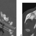
- Alobar: It is the most severe form (Fig. 10B). Complete failure of cleavage, so the falx and interhemispheric fissure are absent, and the thalami and basal ganglia are fused with a holoventricle. This can communicate with a dorsal cyst (expanded tela choroidea). The sulcation/gyration is extremely poor. There are severe facial anomalies (“the face predicts the brain”) with various degrees of midline structural defects, the extreme form being cyclopia.
- Semilobar: There is a single ventricular cavity, with anterior single frontal lobe, whereas the posterior/dorsal portion of the brain (temporo-occipital lobes) is better divided into distinct hemispheres with an intervening fissure and falx. The basal ganglia and thalami are still partially fused. Posteriorly, fibers cross the midline to form an apparent splenium of the corpus callosum. The hippocampi are poorly formed.
- Lobar: There is cortical continuity of the frontal lobes across the midline, with the most anterior part of the interhemispheric fissure present, with a falx. The septum pellucidum is absent with variable formation of the anterior corpus callosum. The face is normal, but in some genetic disorders, there may be a single midline incisor.
- Syntelencephaly: