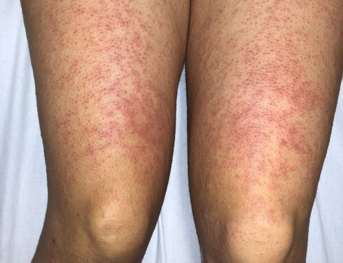
A 37-year-old male with a past medical history of type 2 diabetes presents to the Emergency Department (ED) with a rash. Initial symptoms began one week prior with small spots on the right leg with associated itching and burning. He initially presented to an outside facility where he was diagnosed with an allergic reaction versus scabies and was given a short course of oral steroids and topical permethrin that provided some relief. The rash progressed to bilateral lower extremities prompting re-presentation to the ED. He also reports associated dark urine and nausea.
GI: Abdomen soft, non-tender, non-distended.
MSK: No joint swelling, tenderness, erythema or warmth.
Skin: Numerous scattered bright red palpable purpuric papules and plaques, most concentrated on bilateral lower extremities extending to lower abdomen at the level of the umbilicus.
White blood cell (WBC) count: 14.5 k
Creatinine: 1.1 mg/dL on day of presentation, peaked at 2.2 mg/dL approximately 10 days later.
C-reactive protein (CRP): 32.7 mg/L
Erythrocyte sedimentation rate (ESR): 34 mm/hr
Urinalysis: 3+ protein, 2+ blood, 11-20 RBC, 26-50 WBC, rare bacteria
This is a case of IgA vasculitis, formerly called Henoch-Schonlein purpura or HSP. This diagnosis is suspected when a patient has purpuric skin lesions predominantly on the lower limbs as well as at least one of the following: abdominal pain, joint involvement, renal involvement (proteinuria/hematuria), and biopsy demonstrating IgA deposition. This vasculitis is more commonly seen in children and has a male predominance.
Similar to children with IgA vasculitis, adults presenting with this palpable purpuric rash can have associated joint involvement and GI involvement, though intussusception is less common in the adult population. Renal manifestations are more common in adults with this diagnosis and range from proteinuria and hematuria to renal failure. Our patient initially presented with hematuria/proteinuria and less than two weeks later had a doubled his creatinine. A renal biopsy later confirmed IgA nephropathy.
Take-Home Points
- Consider IgA vasculitis in patients with lower extremity purpuric skin lesions with associated abdominal pain/GI bleed, arthralgia, renal involvement, and/or biopsy confirming IgA deposition.
- In adults with IgA vasculitis, renal involvement is more common and often more severe.
- Ozen S, Pistorio A, Iusan SM, Bakkaloglu A, Herlin T, Brik R, Buoncompagni A, Lazar C, Bilge I, Uziel Y, Rigante D, Cantarini L, Hilario MO, Silva CA, Alegria M, Norambuena X, Belot A, Berkun Y, Estrella AI, Olivieri AN, Alpigiani MG, Rumba I, Sztajnbok F, Tambic-Bukovac L, Breda L, Al-Mayouf S, Mihaylova D, Chasnyk V, Sengler C, Klein-Gitelman M, Djeddi D, Nuno L, Pruunsild C, Brunner J, Kondi A, Pagava K, Pederzoli S, Martini A, Ruperto N; Paediatric Rheumatology International Trials Organisation (PRINTO). EULAR/PRINTO/PRES criteria for Henoch-Schönlein purpura, childhood polyarteritis nodosa, childhood Wegener granulomatosis and childhood Takayasu arteritis: Ankara 2008. Part II: Final classification criteria. Ann Rheum Dis. 2010 May;69(5):798-806. doi: 10.1136/ard.2009.116657. PMID: 20413568.
- Yaseen K, Herlitz LC, Villa-Forte A. IgA Vasculitis in Adults: a Rare yet Challenging Disease. Curr Rheumatol Rep. 2021 Jul 1;23(7):50. doi: 10.1007/s11926-021-01013-x. PMID: 34196893.
Copyright
Images and cases from the Society of Academic Emergency Medicine (SAEM) Clinical Images Exhibit at the 2021 SAEM Annual Meeting | Copyrighted by SAEM 2021 – all rights reserved. View other cases from this Clinical Image Series on ALiEM.
Erin Shufflebarger, MD
Latest posts by Erin Shufflebarger, MD (see all)
- SAEM Clinical Images Series: An Adult with a Lower Extremity Rash - January 30, 2023


