Itch in Scabies—What Do We Know?
 Sascha Ständer
 Sonja Ständer
- 1Department of Dermatology, University of Lübeck, Lübeck, Germany
- 2Department of Dermatology, Center for Chronic Pruritus, University Hospital Münster, Münster, Germany
Scabies is a common parasitic skin infestation characterized by severe itch and a heterogenous clinical presentation. Itch, as the cardinal symptom of scabies, is imposing a high burden on affected patients and is often difficult to manage. Decreased life quality and secondary complications, caused by an itch-related disruption of the epidermal barrier and subsequent superinfections, illustrate the need to treat scabies and to understand the underlying mechanisms of itch in respective patients. This review summarizes available data on itch in scabies with a special focus on the clinical aspects and its underlying pathomechanisms.
Scabies—An Overview
Scabies is a highly contagious ectoparasitic skin infestation caused by Sarcoptes scabiei var. hominis. With a global prevalence of 204 million, scabies poses a remarkable burden on both infected individuals and on the healthcare system regardless of the socioeconomic standard of the respective country (1, 2). More specifically, in high income countries, delayed diagnosis of this neglected disease can lead to waves of institutional outbreaks. In middle-to-low income countries a lack of therapeutic resources often results in secondary scabies-related complications, such as chronic kidney disease since excoriations and the disruption of the epidermal barrier caused by itch may lead to impetigo and to a subsequent dissemination of streptococci to the glomeruli (1, 3). The increased disease-related morbidity and mortality further adds to the patients' burden. Depending on different populations, the prevalence of scabies is ranging from 0.2 to 71.4% (4), with a predominant affection of people living in tropical regions (5). While scabies can occur in every individual, current data reveal a greater susceptibility for the young, old, and, generally, immunocompromised patients (6). Accordingly, Mason et al. reported the highest prevalence of scabies in infants <1 year of age [34.1%, adjusted odds ratio (AOR) compared with adults: 3.6, 95%CI 2.2–6.0] and children aged 1–4 years (25.7%, AOR 2.6, 95%CI 1.7–3.9) in the Solomon Islands (7). Furthermore, the disability-adjusted life-years (DALY) burden was found to be the highest in children 1–4 years-old, eventually decreasing from age 5–24 years and recurrently rising after the age of 70 years in the Global Burden of Disease Study 2015 (5). Furthermore, in low-income countries, disadvantaged populations and children under the age of two bare a greater risk to get infected with scabies (4).
The mite Sarcoptes scabiei is an obligate human parasite that burrows into the epidermis mostly after intense skin to skin contact but also after contact to mites from textiles (i.e., sleeping in a bed with mites). The female mite lays eggs and after approximately 14 days the hatched larvae, and later the nymph, reach adulthood (8), provoking symptoms mostly after 2-5 weeks of latency after the first infestation. After a second infestation, the aforementioned symptoms can occur earlier (~ after 1–2 weeks) due to the immune memory. As scabies mites prefer areas with a higher body temperature and a rather thin stratum corneum, predilection sites are the interdigital spaces of hands and feet, the axillary and periumbilical region, the penis and the perianal skin, while the head and neck are usually spared (exception in infants and old people) (9).
The clinical picture of the disease is caused by the infestation of the mites per se and by an immunologic reaction elicited by the contact toward the by-products of the mites (i.e., saliva, excrements). This delayed type reaction of cellular immunity is clinically apparent as an eczematous morphology with multiple disseminated erythematous papules and vesicles on red skin (Figure 1). Due to an intense pruritus, especially at night, secondary skin lesions, that is, excoriations and scratch marks occur frequently. Secondary superinfections in terms of impetigo can frequently be observed in children.
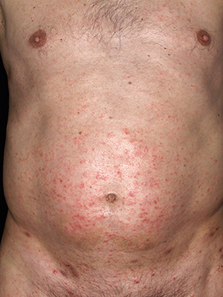
Figure 1. Clinical presentation of a male patient infected with scabies: Multiple erythematous papules in the periumbilical region with several scratch lesions and excoriations.
Scabies crustosa (norvegica), a comparably rare and severe form of scabies with a prevalence of <0.1%, is especially apparent in individuals with an underlying immunosuppression, that is, HIV (10). The localized or generalized hyperkeratotic clinical picture results from a massive mite proliferation and is associated with an increased mortality and a lower grade of itch (10, 11) (Figure 2).
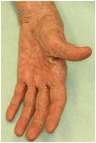
Figure 2. Crusted scabies: Interdigitally located squamous plaques and disseminated erythematous papules.
Diagnosis of scabies is based on a characteristic history (intense pruritus with a nightly deterioration, frequent relapses and refractory to treatment with topical corticosteroids) and the clinical presentation with skin lesions along the predilection sites and typical findings in dermatoscopy and positive microscopy, that is, mites, feces, eggs or mite passages.
Currently, topical treatment with permethrin cream (5%) is considered as the gold standard, leading to a cure in most of the cases (12). Yet, increasing numbers of failures in treatment by permethrin have been recently reported also hinting toward the possibility of increased tolerance of permethrin (when applied under controlled conditions) or possible application errors (13, 14). Successful treatment, however, highly depends on the appropriate application of the cream (whole body from the neck) and concomitant treatment of persons with close contact and basic hygiene measures. Relapses, however, occur frequently; particularly in larger families and groups. Treatment with oral ivermectin has proven to remarkably decrease the prevalence of scabies in larger communities with a good tolerability and efficacy. Recent data is additionally hinting toward the safe and effective use of ivermectin in infants weighing <15 kg (6, 15). Besides these two most commonly used treatments in Europe and the US, various other options are available (16). Sulfur compounds are widely used in Afrika and South America due to good efficacy, however, skin irritations are frequent. Benzyl benzoate is highly antiscabietic but also bares high rates of skin irritation. Both treatment options are commonly used in poorer regions due to their efficacy and low cost. Crotamiton is a topical treatment that shows a good tolerability and is therefore used in children with unsatisfactory success (16).
Clinical Characteristics of Itch in Patients Infected With Scabies
The infestation with scabies poses a high burden on respective patients, often due to an intense unbearable itch. The symptom lasts as long as scabies lasts, however, it might become chronic due to persistence after therapy of the infestation. Thus, scabies should be excluded in cases of persisting pruritus especially with a history of itch in persons with close skin contact (17). In severe cases, the patients even develop papules due to scratching (prurigo), or eczema. Management of chronic itch is often challenging (18) and especially scabies-specific itch is poorly characterized and understood. Itch can deteriorate over the night (nocturnal crescendo). This, however, is not specifically applicable to scabies, as other common skin diseases also display a nighty worsening of itch, that is, atopic dermatitis and psoriasis (19–22). Concerning other clinical symptoms, Brenaut et al. found that heat sensations accompanying the itch were significantly less frequent as compared to the other pruritic skin diseases (19). Sweating and hot water increased the intensity of itch in 73 and 67% of scabies patients, respectively. Scratching was considered pleasurable in 47% of scabies patients as compared to 69 and 65% of patients with atopic dermatitis and psoriasis, respectively. Interestingly, while scratching lesions in patients infected with scabies (63% of patients, p < 0.01) were significantly more frequent as compared to non-atopic eczema, psoriasis and urticaria, patients with scabies revealed the lowest rate of lichenification (8%) compared to 80, 61, and 33% of patients with atopic dermatitis, eczema and psoriasis, respectively. Of note, one major limitation of the study is the depiction of qualitative but not quantitative itch features between the aforementioned dermatosis (19).
The presence of pruritus is reported in most of the patients infected with scabies and the prevalence ranges from 90–99% in current literature (23). Nair et al. found a reported prevalence of pruritus in 99% of 102 adults and a nocturnal aggravation in nearly 80% of the patients leading to sleep disturbances in a prospective, observational cross-sectional study conducted at a tertiary center (24). Similar data regarding the manifestation of itch was reported from a cohort of 323 pediatric patients (25). Here, the overall itch prevalence accounted for 94.5% with a range from 90.3–96.9%. Interestingly, the authors showed that the sensation of itch increases with the age of the pediatric patients characterized in the cohort (25). Itch, however, is more difficult to assess in infant patients and is often displayed as discomfort, crying and an increased irritability what might explain the aforementioned observation.
Comparing classical scabies with scabies crustosa/norvegica, differences in pruritus have been reported, indicating that the itch intensity is lower in patients with crusted scabies. However, still most of the patients with crusted scabies had pruritus to some extend (10, 11). Interestingly, in the crusted scabies cohort characterized by Roberts et al. more than half of the patients bared an identifiable immunosuppressive risk. The authors stated that in patients without respective risks, the development of the crusted clinical appearance might result from an increased tendency to mount a Th2 immune response (10). Furthermore, crusted scabies is reported to affect predominantly individuals with malnutrition, Down's syndrome, the elderly and patients with deficient cognitive abilities or physical debilities who are unable to appropriately depict and to react to itch by scratching (10, 26, 27).
Pathophysiology of Itch in Scabies
Yet, insights into the exact underlying pathomechanisms of itch in scabies remain scant while remarkable progress in understanding itch in principal had been made in the past 20 years (23). The pathophysiology of itch in general includes the direct stimulation of itch-sensory neurons in the skin by epithelial-cell-derived cytokines, that is, IL-33 and thymic stromal lymphopoietin (TSLP) and an indirect stimulation of itch by keratinocyte-derived kalikreins (KLK) like KLK7. Furthermore, the effector cytokines IL-4, IL-9, IL-13, and IL-31 and CXCL10 directly promote itch (28). Regarding the pruritogens IL-4, IL-13, and downstream JAK activation, no studies investigated this in scabies in detail.
However, hypothesis can be derived from the immunologic reaction ongoing in a patient infected with scabies and by using a novel porcine animal model for scabies, thus, deeper insights into the scabies-specific itch can be generated.
The major immune response to the mite infestation includes the innate immune system and the activation of the complement system that, so some extent, can be inhibited by components produced by the mites. Effectors of the immune response include activated mast cells, immunoglobulin E (IgE), eosinophils, and non-histaminergic effectors like PAR2 and IL-31. While in the classical non-crusted scabies, a Th1 mediated immune response plays a predominant role, a Th2 immune response seems to be more important in the pathogenesis of crusted scabies (29).
Albeit immunohistochemical analysis, using a basophil-specific BB1 antibody, revealed numerous basophils infiltrating lesional scabies-infested skin, the pathogenetic significance of this observation remains unclear and requires further investigations (30, 31).
Current hypothesis regarding the pathophysiology of itch in scabies can be subdivided into the direct action of the scabies mite and the immune response toward the mite itself (23).
Mite components can directly lead to an activation of the Toll-like receptor (TLR) pathway with a subsequent activation of TLR 3, 4, and 7 that are expressed on primary sensory neurons (32, 33). Mite feces contain proteases that can lead to an activation of protease-activated receptor 2 (34). The close interaction between the mites and the keratinocytes can lead to the release of protease activating protease-activated prurireceptors (23, 35). Furthermore, mite components that can be recognized as antigens and show a similar structure to antigens of the house dust mite, can induce an IgE-mediated mast cell activation with an aggravation of itch mediated by the degranulation of histamine, tumor necrosis factor (TNF) alpha and tryptase with a subsequent activation of histaminergic H1 and H4 prurireceptors and protease-activated prurireceptors by tryptase (36, 37). The itch sensation can further be enhanced by a release of leukotriens and prostaglandins implicated my macrophages (23).
In the Th1-mediated immune response that is primarily present in classical non-crusted scabies, a release of INF-gamma and interleukin(IL)-2 lead to an activation of cytokine prurireceptors, whereas an up-regulation of the Th2-mediated immune response in crusted scabies elevates eosinophils, IgE-activated mast cells and enhances the activation of cytokine prurireceptors by IL-31 (23, 37). Recently, it was shown that increased IL-31 levels from murine peritoneal macrophages were induced by an overexpression of thymic stromal lymphopoietin and periostin in an experimental mouse model (38). However, the relevance of this non-histaminergic pathway in the scabies itch is yet to be further investigated as compared to the histaminergic pathway.
In the recent years, a porcine model for scabies was established facilitating the investigation of specific scabies-related questions (39–41). Recently, Sanders et al. investigated the potential mechanisms of scabies itch and found that non-histaminergic mediators of pruritus were significantly elevated in the skin of pigs experimentally infested with scabies as well as in human skin as compared to non-infected healthy controls (42). Accordingly, a significant upregulation of TRPV1, TRPA1, and PAR-2 expression in the epidermis and an increase of tryptase+ cells around the dermal-epidermal junction was found in both porcine and human scabies-infected skin. These data suggest that the non-histaminergic mediators might play an important role in scabies itch and might potentially serve as therapeutic targets. Furthermore, the similar results from human and pig skin biopsies indicate that the porcine model might serve as suitable animal model to investigate the scabies-specific itch in future experiments. Slight variances in the data might result from different biopsy sites, different durations of the disease, scratching and general differences between the species (42).
Complications Linked to Itch in Scabies
Usually, secondary complications and morbidity of individuals infected with scabies are discussed as a direct consequence of the infestation with scabies. However, it is more accurate to refer to subsequent complications due to massive itch caused by an immune response toward the mites and the direct interaction of complement inhibitors produced by the mites and complement pathways in the skin enabling bacteria to grow more easily. Furthermore, severe itch and subsequent scratching leads to a disruption of the epidermal skin barrier (43) and thus to a skin more susceptible for bacterial skin infections. Due to the ability of the scabies mite to interfere with the human complement system by blocking all three complement initiation pathways and leading to decreased neutrophil functions, staphylococcal and streptococcal growth is promoted (44–50). A study performed using the porcine animal model provided evidence that the skin microbiome is changed due to the scabies infestation enabling the growth of opportunistic pathogens (51). In literature, impetigo is described as the most frequent complication of scabies-related itch. It commonly affects children and individuals living under crowded conditions in tropical regions (4, 52, 53). Excoriated deep skin lesions harboring bacteria, most frequently Staphylococcus aureus and group A Streprococcus (AGU), can further lead to a hematogenic dissemination with subsequent complications involving other organs. Interestingly, scratching lesions appear more frequently in patients infested with scabies as compared to other itching dermatosis i.e. eczema, psoriasis and uritcaria (19). The bacterial superinfection can lead to local infections (i.e., erysipelas, cellulitis, abscesses, staphyloderma) with/or without systemic affection and in the worst case to a sepsis. Post-streptococcal complications can affect the kidneys (glomerulonephritis), the heart (rheumatic heart disease) and the joints (acute rheumatic fever) and pose a high burden on respective patients and the healthcare system (54–57). Patients infected with scabies, thus, suffer from an impaired life quality that is directly linked to the severity of itch.
Conclusion
Scabies is a common and neglected skin infestation characterized by severe itch and a heterogenous clinical picture. Itch in scabies can be caused by direct mite actions and by a resulting immune response toward the mites. Recent data from the porcine animal model hint toward an important role of non-histaminergic itch mediators, that is, TRPV1, TRPA1, PAR-2, and tryptase+ cells. The exact pathomechanism of scabies-specific itch remains yet to be further investigated.
Author Contributions
All authors contributed to the article and approved the submitted version.
Conflict of Interest
The authors declare that the research was conducted in the absence of any commercial or financial relationships that could be construed as a potential conflict of interest.
References
1. Thomas C, Coates SJ, Engelman D, Chosidow O, Chang AY. Ectoparasites: scabies. J Am Acad Dermatol. (2020) 82:533–48. doi: 10.1016/j.jaad.2019.05.109
2. GBD 2015 Disease and Injury Incidence and Prevalence Collaborators. Global, regional, and national incidence, prevalence, and years lived with disability for 310 diseases and injuries, 1990–2015: a systematic analysis for the Global Burden of Disease Study 2015. Lancet. (2016) 388:1545–602. doi: 10.1016/S0140-6736(16)31678-6
3. Chung S-D, Wang K-H, Huang C-C, Lin H-C. Scabies increased the risk of chronic kidney disease: a 5-year follow-up study. J Eur Acad Dermatol Venereol. (2014) 28:286–92. doi: 10.1111/jdv.12099
4. Romani L, Steer AC, Whitfeld MJ, Kaldor JM. Prevalence of scabies and impetigo worldwide: a systematic review. Lancet Infect Dis. (2015) 15:960–7. doi: 10.1016/S1473-3099(15)00132-2
5. Karimkhani C, Colombara DV, Drucker AM, Norton S, Hay R, Engelmann D, et al. The global burden of scabies: a cross-sectional analysis from the Global Burden of Disease Study 2015. Lancet Infect Dis. (2017) 17:1247–54. doi: 10.1016/S1473-3099(17)30483-8
6. Ständer S, Kirschstein DJ, Kohl-Sobania M, Zillikens D, Ludwig RJ, Anemüller W. Effectiveness and adverse events of ivermectin treatment for scabies in 30 infant patients: report from a German single centre. J Eur Acad Dermatol Venereol. (2020) 34:e736–7. doi: 10.1111/jdv.16554
7. Mason DS, Marks M, Sokana O, Solomon A, Mabey DC, Romani L, et al. The prevalence of scabies and impetigo in the Solomon Islands: a population-based survey. PLoS Negl Trop Dis. (2016) 10:e0004803. doi: 10.1371/journal.pntd.0004803
8. Arlian LG, Morgan MS. A review of Sarcoptes scabiei: past, present and future. Parasit Vectors. (2017) 10:297. doi: 10.1186/s13071-017-2234-1
9. Sunderkötter C, Mayser P, Fölster-Holst R, Maier WA, Kampen H, Hamm H. Scabies. J Dtsch Dermatol Ges. (2007) 5:424–30. doi: 10.1111/j.1610-0387.2007.06298.x
10. Roberts LJ, Huffam SE, Walton SF, Currie BJ. Crusted scabies: clinical and immunological findings in seventy-eight patients and a review of the literature. J Infect. (2005) 50:375–81. doi: 10.1016/j.jinf.2004.08.033
11. Karthikeyan K. Crusted scabies. Indian J Dermatol Venereol Leprol. (2009) 75:340–7. doi: 10.4103/0378-6323.53128
12. Salavastru CM, Chosidow O, Boffa MJ, Janier M, Tiplica GS. European guideline for the management of scabies. J Eur Acad Dermatol Venereol. (2017) 31:1248–53. doi: 10.1111/jdv.14351
14. Hackenberg B, Horváth ON, Petachti M, Schult R, Yenigün N, Bannenberg P. Scabies therapy in Germany : Results of a nationwide survey with a special focus on the efficacy of first-line therapy with permethrin. Hautarzt. (2020) 71:374–9. doi: 10.1007/s00105-020-04561-y
15. Levy M, Martin L, Bursztejn A-C, Chiaverini C, Miquel J, Mahé E, et al. Ivermectin safety in infants and children under 15 kg treated for scabies: a multicentric observational study. Br J Dermatol. (2019) 182:1003–6. doi: 10.1111/bjd.18369
16. Chandler DJ, Fuller LC. A Review of scabies: an infestation more than skin deep. DRM. (2019) 235:79–90. doi: 10.1159/000495290
17. Hengge UR, Currie BJ, Jäger G, Lupi O, Schwartz RA. Scabies: a ubiquitous neglected skin disease. Lancet Infect Dis. (2006) 6:769–79. doi: 10.1016/S1473-3099(06)70654-5
18. Pereira MP, Zeidler C, Storck M, Agelopoulos K, Philipp-Dormston W, Ständer S, et al. Challenges in clinical research and care in pruritus. Acta Derm Venereol. (2020) 100:adv00028. doi: 10.2340/00015555-3348
19. Brenaut E, Garlantezec R, Talour K, Misery L. Itch characteristics in five dermatoses: non-atopic eczema, atopic dermatitis, urticaria, psoriasis and scabies. Acta Derm Venereol. (2013) 93:573–4. doi: 10.2340/00015555-1599
20. Li JC, Fishbein A, Singam V, Patel KR, Zee PC, Attarian H, et al. Sleep disturbance and sleep-related impairment in adults with atopic dermatitis: a cross-sectional study. Dermatitis. (2018) 29:270–7. doi: 10.1097/DER.0000000000000401
21. Kimball AB, Edson-Heredia E, Zhu B, Guo J, Maeda-Chubachi T, Shen W, et al. Understanding the relationship between pruritus severity and work productivity in patients with moderate-to-severe psoriasis: sleep problems are a mediating factor. J Drugs Dermatol. (2016) 15:183–8.
22. Lavery MJ, Stull C, Nattkemper LA, Sanders KM, Lee H, Valdes-Rodriguez R, et al. Nocturnal pruritus: prevalence, characteristics, and impact on ItchyQoL in a chronic itch population. Acta Derm Venereol. (2017) 97:513–5. doi: 10.2340/00015555-2560
23. Jannic A, Bernigaud C, Brenaut E, Chosidow O. Scabies itch. Dermatol Clin. (2018) 36:301–8. doi: 10.1016/j.det.2018.02.009
24. Nair PA, Vora RV, Jivani NB, Gandhi SS. A study of clinical profile and quality of life in patients with scabies at a rural tertiary care centre. J Clin Diagn Res. (2016) 10:WC01–5. doi: 10.7860/JCDR/2016/20938.8703
25. Boralevi F, Diallo A, Miquel J, Guerin-Moreau M, Bessis D, Chiavérini C, et al. Clinical phenotype of scabies by age. Pediatrics. (2014) 133:e910–6. doi: 10.1542/peds.2013-2880
26. Chosidow O. Scabies and pediculosis: neglected diseases to highlight. Clin Microbiol Infect. (2012) 18:311–2. doi: 10.1111/j.1469-0691.2012.03791.x
27. Sullivan JR, Watt G, Barker B. Successful use of ivermectin in the treatment of endemic scabies in a nursing home. Australas J Dermatol. (1997) 38:137–40. doi: 10.1111/j.1440-0960.1997.tb01130.x
28. Wang F, Kim BS. Itch: a paradigm of neuroimmune crosstalk. Immunity. (2020) 52:753–66. doi: 10.1016/j.immuni.2020.04.008
29. Bhat SA, Mounsey KE, Liu X, Walton SF. Host immune responses to the itch mite, Sarcoptes scabiei, in humans. Parasit Vectors. (2017) 10:385. doi: 10.1186/s13071-017-2320-4
30. Ito Y, Satoh T, Takayama K, Miyagishi C, Walls AF, Yokozeki H. Basophil recruitment and activation in inflammatory skin diseases. Allergy. (2011) 66:1107–13. doi: 10.1111/j.1398-9995.2011.02570.x
31. Hashimoto T, Rosen JD, Sanders KM, Yosipovitch G. Possible roles of basophils in chronic itch. Exp Dermatol. (2019) 28:1373–9. doi: 10.1111/exd.13705
32. He R, Gu X, Lai W, Peng X, Yang G. Transcriptome-microRNA analysis of Sarcoptes scabiei and host immune response. PLoS ONE. (2017) 12:e0177733. doi: 10.1371/journal.pone.0177733
33. Taves S, Ji R-R. Itch control by toll-like receptors. Handb Exp Pharmacol. (2015) 226:135–50. doi: 10.1007/978-3-662-44605-8_7
34. Lavery MJ, Stull C, Kinney MO, Yosipovitch G. Nocturnal pruritus: the battle for a peaceful night's sleep. Int J Mol Sci. (2016) 17:425. doi: 10.3390/ijms17030425
35. Raap U, Ständer S, Metz M. Pathophysiology of itch and new treatments. Curr Opin Allergy Clin Immunol. (2011) 11:420–7. doi: 10.1097/ACI.0b013e32834a41c2
36. Morgan MS, Rider SD, Arlian LG. Identification of antigenic Sarcoptes scabiei proteins for use in a diagnostic test and of non-antigenic proteins that may be immunomodulatory. PLoS Negl Trop Dis. (2017) 11:e0005669. doi: 10.1371/journal.pntd.0005669
37. Azimi E, Xia J, Lerner EA. Peripheral mechanisms of itch. Curr Probl Dermatol. (2016) 50:18–23. doi: 10.1159/000446012
38. Hashimoto T, Satoh T, Yokozeki H. Pruritus in ordinary scabies: IL-31 from macrophages induced by overexpression of thymic stromal lymphopoietin and periostin. Allergy. (2019) 74:1727–37. doi: 10.1111/all.13870
39. Bernigaud C, Fang F, Fischer K, Lespine A, Aho LS, Dreau D, et al. Preclinical study of single-dose moxidectin, a new oral treatment for scabies: efficacy, safety, and pharmacokinetics compared to two-dose ivermectin in a porcine model. PLoS Negl Trop Dis. (2016) 10:e0005030. doi: 10.1371/journal.pntd.0005030
40. Bernigaud C, Fang F, Fischer K, Lespine A, Aho LS, Mullins AJ, et al. Efficacy and pharmacokinetics evaluation of a single oral dose of afoxolaner against sarcoptes scabiei in the porcine scabies model for human infestation. Antimicrob Agents Chemother. (2018) 62:e02334–17. doi: 10.1128/AAC.02334-17
41. Mounsey K, Ho M-F, Kelly A, Willis C, Pasay C, Kemp DJ, et al. A tractable experimental model for study of human and animal scabies. PLoS Negl Trop Dis. (2010) 4:e756. doi: 10.1371/journal.pntd.0000756
42. Sanders KM, Nattkemper LA, Rosen JD, Andersen HH, Hsiang J, Romanelli P, et al. Non-Histaminergic itch mediators elevated in the skin of a porcine model of scabies and of human scabies patients. J Invest Dermatol. (2019) 139:971–3. doi: 10.1016/j.jid.2018.09.032
43. Yosipovitch G, Misery L, Proksch E, Metz M, Ständer S, Schmelz M. Skin barrier damage and itch: review of mechanisms, topical management and future directions. Acta Derm Venereol. (2019) 99:1201–9. doi: 10.2340/00015555-3296
44. Swe PM, Christian LD, Lu HC, Sriprakash KS, Fischer K. Complement inhibition by Sarcoptes scabiei protects Streptococcus pyogenes—an in vitro study to unravel the molecular mechanisms behind the poorly understood predilection of S. pyogenes to infect mite-induced skin lesions. PLoS Negl Trop Dis. (2017) 11:e0005437. doi: 10.1371/journal.pntd.0005437
45. Swe PM, Reynolds SL, Fischer K. Parasitic scabies mites and associated bacteria joining forces against host complement defence. Parasite Immunol. (2014) 36:585–93. doi: 10.1111/pim.12133
46. Swe PM, Fischer K. A scabies mite serpin interferes with complement-mediated neutrophil functions and promotes staphylococcal growth. PLoS Negl Trop Dis. (2014) 8:e2928. doi: 10.1371/journal.pntd.0002928
47. Mika A, Reynolds SL, Mohlin FC, Willis C, Swe PM, Pickering DA, et al. Novel scabies mite serpins inhibit the three pathways of the human complement system. PLoS ONE. (2012) 7:e40489. doi: 10.1371/journal.pone.0040489
48. Mika A, Reynolds SL, Pickering D, McMillan D, Sriprakash K, Kemp DJ, et al. Complement inhibitors from scabies mites promote streptococcal growth–a novel mechanism in infected epidermis? PLoS Negl Trop Dis. (2012) 6:e1563. doi: 10.1371/journal.pntd.0001563
49. Bergström FC, Reynolds S, Johnstone M, Pike RN, Buckle AM, Kemp DJ, et al. Scabies mite inactivated serine protease paralogs inhibit the human complement system. J Immunol. (2009) 182:7809–17. doi: 10.4049/jimmunol.0804205
50. Reynolds SL, Pike RN, Mika A, Blom AM, Hofmann A, Wijeyewickrema L, et al. Scabies mite inactive serine proteases are potent inhibitors of the human complement lectin pathway. PLoS Negl Trop Dis. (2014) 8:e2872. doi: 10.1371/journal.pntd.0002872
51. Swe PM, Zakrzewski M, Kelly A, Krause L, Fischer K. Scabies mites alter the skin microbiome and promote growth of opportunistic pathogens in a porcine model. PLoS Negl Trop Dis. (2014) 8:e2897. doi: 10.1371/journal.pntd.0002897
52. Romani L, Marks M, Sokana O, Nasi T, Kamoriki B, Cordell B, et al. Efficacy of mass drug administration with ivermectin for control of scabies and impetigo, with coadministration of azithromycin: a single-arm community intervention trial. Lancet Infect Dis. (2019) 19:510–8. doi: 10.1016/S1473-3099(18)30790-4
53. Reid HF, Birju B, Holder Y, Hospedales J, Poon-King T. Epidemic scabies in four Caribbean islands, 1981-1988. Trans R Soc Trop Med Hyg. (1990) 84:298–300. doi: 10.1016/0035-9203(90)90296-Q
54. Streeton CL, Hanna JN, Messer RD, Merianos A. An epidemic of acute post-streptococcal glomerulonephritis among aboriginal children. J Paediatr Child Health. (1995) 31:245–8. doi: 10.1111/j.1440-1754.1995.tb00795.x
55. Lawrence G, Leafasia J, Sheridan J, Hills S, Wate J, Wate C, et al. Control of scabies, skin sores and haematuria in children in the Solomon Islands: another role for ivermectin. Bull World Health Organ. (2005)83:34–42.
56. Steer AC, Jenney AWJ, Kado J, Batzloff MR, La Vincente S, Waqatakirewa L, et al. High burden of impetigo and scabies in a tropical country. PLoS Negl Trop Dis. (2009) 3:e467. doi: 10.1371/journal.pntd.0000467
Keywords: scabies, itch, pruritus, itch mediators, itch and its pathways
Citation: Ständer S and Ständer S (2021) Itch in Scabies—What Do We Know? Front. Med. 8:628392. doi: 10.3389/fmed.2021.628392
Received: 11 November 2020; Accepted: 05 January 2021;
Published: 01 February 2021.
Edited by:
Peter Wolf, Medical University of Graz, AustriaReviewed by:
William Faber, Amsterdam University Medical Center (UMC), NetherlandsAlessandra Handisurya, Medical University of Vienna, Austria
Copyright © 2021 Ständer and Ständer. This is an open-access article distributed under the terms of the Creative Commons Attribution License (CC BY). The use, distribution or reproduction in other forums is permitted, provided the original author(s) and the copyright owner(s) are credited and that the original publication in this journal is cited, in accordance with accepted academic practice. No use, distribution or reproduction is permitted which does not comply with these terms.
*Correspondence: Sascha Ständer, sascha.staender@uksh.de