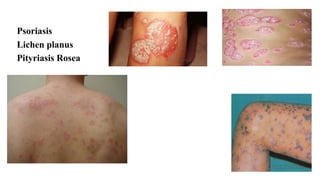
3. Psoriasis
An inflammatory disease
Clinical signs: well-circumscribed, erythematous papules and plaques covered with
silvery scales
Cause: unclear, but seems to involve the immune system
Triggers: trauma, infection, and certain drugs
Symptoms: occasional mild itching, but cosmetic implications may be major. Some
people develop severe disease with painful arthritis
Diagnosis: based on appearance and distribution of lesions
Treatment: emollients, vitamin D analogs, retinoids, tar, anthralin, corticosteroids,
phototherapy, and, when severe, methotrexate, retinoids, immunomodulatory agents
(biologics), or immunosuppressants
Pathophysiology: hyperproliferation of epidermal keratinocytes combined with
inflammation of the epidermis and dermis
Epidemiology: affects about 1 to 5% of the population worldwide
4. Incidence
• Psoriasis is universal in occurrence
• In the United States, psoriasis affects about 2 percent of the population ( 150,000 newly
diagnosed cases per year) affected
• 0.97 percent in South America
• 1.3 percent in Germany
• 1.6 percent in Great Britain,
• 1.7 percent in Denmark
• 2.3 percent in Sweden
• Psoriasis is rare in West African and North American blacks
• The incidence of the disease is also low in Japanese and Eskimos
• Psoriasis is nearly absent in North American Indians, and in an examination of 26,000 South
American Indians, not a single case was seen
• Psoriasis is equally common in males and females
5. Age of Onset
• The onset of psoriasis constitutes a lifelong threat
• Most patients develop the initial lesions of psoriasis in the third decade of life
• A first peak incidence is at 22.5 years of age, a second peak of onset around age 55
• In children, the mean age of onset was 8.1 years
• An early onset (before age 15) predicts more severe disease
• The earlier the onset, the greater is the probability of a positive family history of psoriasis
Mode of Inheritance
• There is a genetic predisposition to psoriasis
• When one parent had psoriasis, psoriasis also developed in 8.1 percent of the offspring
• When both parents had psoriasis this value increased to 41 percent
• The HLA types most frequently reported to be associated with psoriasis are HLA-B13, HLA-
Bw57, HLA-Cw6, and HLA-DR7
6. Lesions of psoriasis show four prominent features:
• they are sharply demarcated with clear-cut borders
• the surface consists of noncoherent silvery scales
• under the scale the skin has a glossy, homogeneous erythema
• there is an Auspitz sign
The size of a single lesion varies from a pinpoint to plaques that cover large areas of
the body.
The Auspitz phenomenon in its three phases:
A. Scratching generates silvery-opaque scale
B. Further scratching leads to removal of the scale, and a glossy area is visible
C. Further scratching produces blood droplets.
In addition to the Auspitz sign, the Koebner phenomenon can be elicited in
approximately 20 percent of patients.
7. Koebner phenomenon
After nonspecific irritation, psoriatic lesions develop in
areas where they were not present previously
The Auspitz sign is a specific feature of the
erythrosquamous lesion of psoriasis. It is noted when the
hyperkeratotic scales are mechanically removed from a
psoriatic plaque by scraping. Within a few seconds after
mechanical removal of the scale, small blood droplets
appear on the shiny erythematous surface
8. Psoriasis
Etiology: (Recent theory) T- cell mediated immune stimulation of epidermal
keratinocytes; certain genes and HLA antigens (Cw6, B13, B17) are associated
with psoriasis
Triggers • Injury (Koebner phenomenon)
• Sunburn
• HIV
• β-Hemolytic streptococcal infection
• Drugs (especially β-blockers, chloroquine, lithium, ACE inhibitors,
indomethacin, terbinafine, and interferon alfa)
• Emotional stress
• Alcohol consumption
10. Psoriasis
Symptoms and Signs
• Lesions are asymptomatic or pruritic
• Localized on the scalp, extensor surfaces of the elbows and knees, sacrum,
buttocks, and penis. The nails, eyebrows, axillae, umbilicus, and perianal
region may also be involved
• Plaque psoriasis (psoriasis vulgaris or chronic plaque psoriasis) is the most
common pattern
• Lesions are discrete, erythematous papules or plaques covered with thick,
silvery, shiny scales. Lesions appear gradually and remit and recur either
spontaneously or with appearance and resolution of triggers
• Arthritis develops in 5 to 30% of patients and can be disabling; joint
destruction may ultimately occur
• Psoriasis is affect a patient's quality of life
11. Psoriasis
Diagnosis
• Clinical evaluation
• Rarely biopsy
Disease is graded as mild, moderate, or severe based on the body surface area
affected and how the lesions affect patients' quality of life (PASI).
Treatment
• Topical treatments (emollients, salicylic acid, coal tar, anthralin,
corticosteroids, vitamin D3 analogs topical retinoids, topical calcineurin
inhibitors)
• Systemic treatments (methotrexate, oral retinoids, and oral calcineurin
inhibitors, immune-suppressants, immunomodulatory agents (biologics))
• Ultraviolet (UV) light therapy
12. NAIL Psoriasis
Nail changes are frequent in psoriasis
1. Pits are evident within the nail plate. This
morphologic pattern apparently is due to defective
keratinization of the dorsal side of the proximal nail
fold
2. Yellowish macules beneath the nail plate often
extend distally toward the hyponychium. This
morphologic pattern appears to be caused by
psoriatic processes located in the nail bed
3. Severe onychodystrophy results in yellowish
keratinous material. This morphologic pattern is
believed to be secondary to psoriasis involving the
nail matrix
17. Guttate psoriasis
• presents as small (0.5–1.5 cm in diameter)
lesions over the upper trunk and proximal
extremities
• Is found frequently in young adults
• Streptococcal throat infection frequently
precedes the onset or flare of guttate
psoriasis
• This process usually signals an acute
exacerbation of disease
• Predisposing factors for such an event are
bacterial infection, aggressive local therapy,
or withdrawal of systemic glucocorticoids
18. Inverse psoriasis
The skin in the intertriginous areas is
highly erythematous, and typical
scaling is lacking
19. Pustular Psoriasis
• Generalized form: pustular psoriasis (von
Zumbusch)
• Localized variant, confined to the palms and
soles, known as pustulosis palmaris et
plantaris
• In rare instances in psoriasis of the plaque
type or guttate psoriasis, pustules may
develop after acute relapses (psoriasis with
pustules).
20. Erythrodermic psoriasis
• Psoriatic erythroderma represents the generalized
form of the disease that affects all body sites,
including the face, hands, feet, nails, trunk, and
extremities
• Although all the symptoms of psoriasis are present,
erythema is the most prominent feature, and scaling
usually is less severe compared with chronic
stationary Psoriasis
• Psoriatic erythroderma may have different degrees
of disease activity, presenting suddenly as a
generalized erythema or evolving gradually from
chronic plaque psoriasis into a generalized
exfoliative phase.
21. Plaque Psoriasis
• Red scaly lesions persist for months to years
• There is constant production of large
amounts of scale with little alteration in
shape or distribution of individual plaques
• Areas of predilection are the elbows, the
knees, the scalp and, in particular, the
retroauricular region, the lumbar area, and
the umbilicus.
• Single small lesions may become confluent,
forming plaques
23. Lichen planus (LP)
A recurrent, pruritic, inflammatory eruption characterized by small, discrete,
polygonal, flat-topped, violaceous papules that may coalesce into rough scaly
patches, often accompanied by oral lesions
Diagnosis: clinical avaluation
skin biopsy
Treatment: topical or intralesional corticosteroids. Severe cases may require
phototherapy or systemic immuno-suppressants
• Etiology: T cell-mediated autoimmune reaction against basal epithelial
keratinocytes in people with genetic predisposition
• Drugs (especially β-blockers, NSAIDs, ACE inhibitors, sulfonylureas, gold,
antimalarial drugs, penicillamine, and thiazides) can cause LP and may have
a pattern that is more eczematous
• Associations with hepatitis C-induced liver insufficiency, other forms of
hepatitis have been reported
24. Symptoms and Signs
• pruritic, purple, polygonal, flat-topped papules and plaques, initially 2 to 4
mm in diameter, with angular borders, a violaceous color, and a distinct sheen
in cross-lighting, symmetrically distributed, on the flexor surfaces of the
wrists, legs, trunk, glans penis, and oral and vaginal mucosae. The face is
rarely involved
• Onset may be abrupt or gradual, new papules may appear at sites of minor
skin injury (Koebner phenomenon), such as a superficial scratch. Lesions
may coalesce, becoming hyperpigmented, atrophic, hyperkeratotic
(hypertrophic LP), or vesiculo bullous, patchy scarring alopecia (lichen
planopilaris) may occur.
• Nails are involved in up to 10% of cases. Findings vary in intensity with nail
bed discoloration, longitudinal ridging and lateral thinning, and complete loss
of the nail matrix and nail, with scarring of the proximal nail fold onto the
nail bed (pterygium formation)
26. Oral Mucosa LP
• The oral mucosa is involved in about 50% of cases
• Reticulated, lacy, bluish-white, linear lesions (Wickham's striae) on the
buccal mucosae and tongue margins and gingival mucosae in edentulous
areas may also be affected
• An erosive form of LP may occur in which the patient develops shallow,
often painful, recurrent oral ulcers, which, if long-standing, rarely become
cancerous
• Vulvar and vaginal mucosae are often involved
• Up to 50% of women with oral mucosal findings have undiagnosed vulvar
LP. In men, genital involvement is common, especially of the glans penis.
• Oral or vaginal LP may resemble leukoplakia
• The oral lesions must also be distinguished from candidiasis, carcinoma,
aphthous ulcers, pemphigus, cicatricial pemphigoid, and chronic erythema
multiforme
27. Lichen Planus
• arranged in a network pattern
(Wickham’s striae) with erythema
of the surrounding mucosa
• White patches, erythematous erosions,
and ulcers may also occur
• The white lesions are not painful, but the
erosions and ulcers are usually painful
• Lichen planus almost always has multiple
lesions bilaterally, with the buccal mucosa
commonly involved. Oral lesions may occur with or
without skin lesions.
• An incisional biopsy is required for definitive diagnosis. Asymptomatic lesions require no
treatment other than inspection during annual dental visits. Topical and/or systemic
corticosteroids will almost always control, but not cure, painful erosions and ulcers of
lichen planus. Candidal overgrowth (candidosis) is common in patients with lichen planus
and should be treated with antifungal medications.
28. Prognosis and treatment
Prognosis: Many cases resolve without intervention, presumably because the
provocative agent is no longer present. Sometimes treatment of a previously
occult infection, such as a dental abscess, results in resolution.
Vulvovaginal LP may be chronic and refractory to therapy, causing decreased
quality of life.
Treatment
• Topical treatments
• Systemic treatments
• Sometimes light therapy
• Asymptomatic LP does not require treatment
• Drugs suspected of triggering LP should be stopped
High-potency ointments or creams used on the thicker lesions on the extremities;
lower-potency drugs may be used on the face, groin, and axillae
Oral corticosteroids, oral retinoids, griseofulvin, cyclosporine, light therapy may
be used for severe cases
29. Treatment of erosive ulcers
• Viscous lidocaine may help relieve symptoms of erosive ulcers.
• Tacrolimus 0.1% ointment applied twice daily may induce lasting remission
Other treatment options include topical (in an adhesive base), intralesional,
and systemic corticosteroids
• Erosive oral LP may respond to oral dapsone or cyclosporine. Cyclosporine
rinses also may be helpful
• Hydroxychloroquine, azathioprine, systemic cyclosporine, and topical
tretinoin may also be useful
• As with any disease with so many therapies, individual drugs have not been
uniformly successful.
32. Herpes Simplex
Vesicles and ulcers anywhere on oral mucosa, the pharynx, lips and perioral skin. The gingiva
is typically enlarged and erythematous. The lesions are painful, making it difficult to eat and
drink. The lesions resolve spontaneously, usually within 10-14 days
The best documented causes of recurrent herpetic lesions* are ultraviolet radiation
mechanical trauma, and immunosuppression.
Recurrent herpes has vesicles and ulcers occurring on keratinized mucosal surfaces. The
lesions are grouped in a tight cluster
Often a sudden prodrome of pain, tingling, or numbness precedes or crops
Oral lesions may occur as vesicles which rupture to form non-painful ulcers
Treatment is usually supportive and symptomatic in immunocompetent children. Antiviral
medications, such as acyclovir, famciclovir, and valacyclovir, are useful in
immunocompromised patients, adults, and infants
A vaccine for varicella is available. It appears to be highly effective, but the duration of
immunity is not known.
34. Pityriasis Rosea
An inflammatory disease characterized by diffuse, scaling papules or
Plaques. Treatment is usually unnecessary
The cause:Viral infection (human herpesviruses 6 and 7?), Drugs may cause a PR-like reaction.
Symptoms and Signs
A single, primary, 2- to 10-cm herald patch that appears on the trunk or proximal limbs and
follows within 7 to 14 days by a general centripetal eruption of 0.5- to 2-cm rose- or fawn-colored
oval papules and plaques with collarette and Christmas tree-like distribution on the back
Diagnosis
• Clinical evaluation
Serologic testing for syphilis.
Treatment
• Antipruritic therapy
the eruption usually remits within 5 wk and recurrence is rare.
Artificial or natural sunlight may hasten resolution.