Copyright
©The Author(s) 2022.
World J Clin Cases. May 26, 2022; 10(15): 4949-4956
Published online May 26, 2022. doi: 10.12998/wjcc.v10.i15.4949
Published online May 26, 2022. doi: 10.12998/wjcc.v10.i15.4949
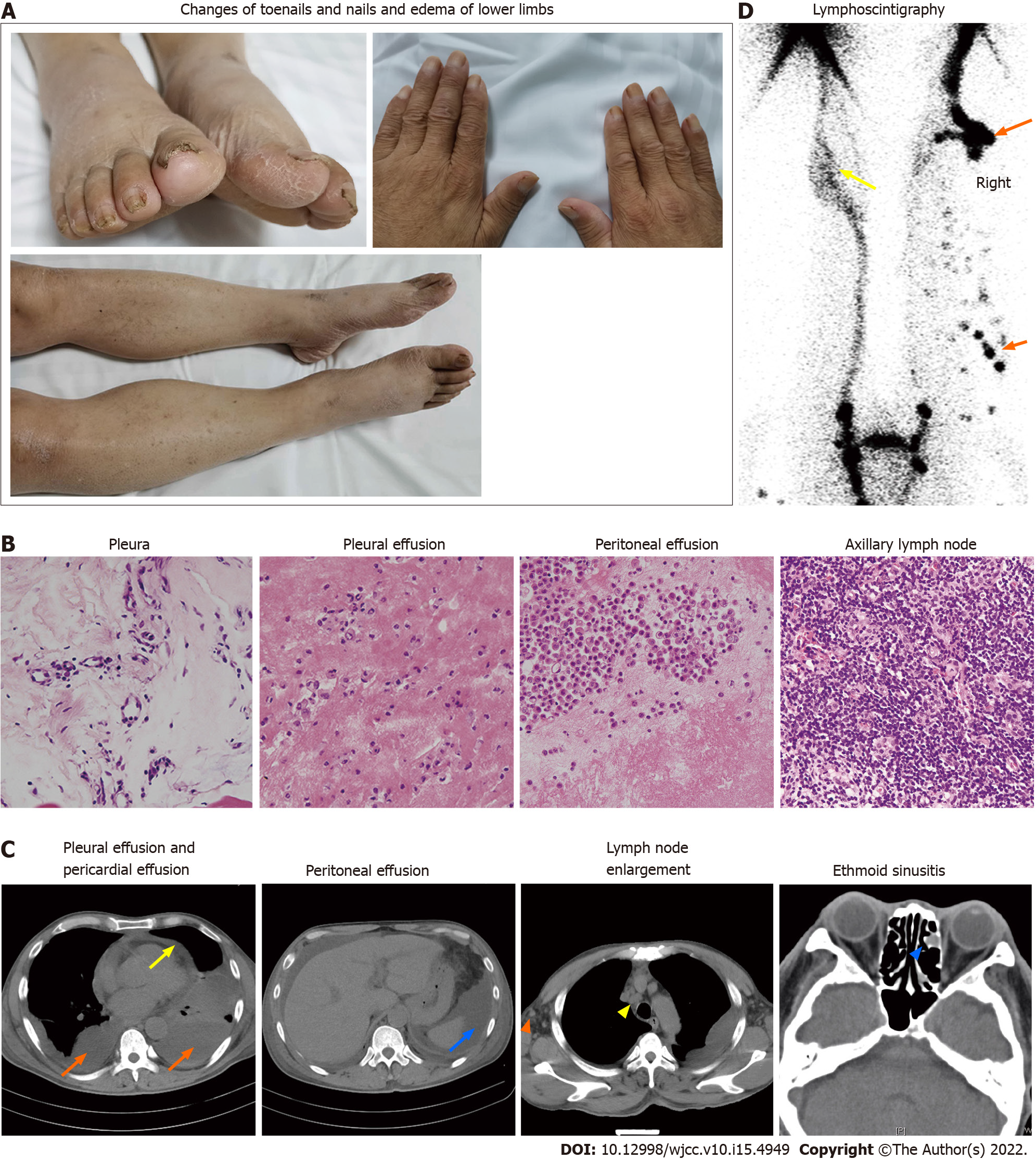
Figure 1 Clinical findings and laboratory examination of the patient.
A: Nails and toenails were thickened and yellow, with increased curvature. Edema was found in both lower limbs, especially in the right side; B: No tumor cells or granulomatous structures were found in hematoxylin and eosin staining images (400×) of pleura, pleural effusion, peritoneal effusion and axillary lymph nodes; C: Computed tomography (CT) images showed bilateral pleural effusions(orange arrows), pericardial effusions (yellow arrow), and peritoneal effusions (blue arrow); positron emission tomography (PET)/CT images showed enlarged axillary (orange triangle) and mediastinal (yellow triangle) lymph nodes and ethmoid sinusitis (blue triangle); D: Radionuclide lymphoscintigraphy showed that lymphatic vessels of the right lower limb were interrupted, the imaging agent (99Tc-antimony sulfide colloid) was accumulated in the subcutaneous tissue (orange arrows) and collateral circulation appeared in the lymphatic vessels below the left knee (yellow arrows).
- Citation: Zhang YN, Wang MH, Yu WC, Cheng W, Cong JP, Huang XP, Wang FF. Yellow nail syndrome accompanied by minimal-change nephrotic syndrome: A case report. World J Clin Cases 2022; 10(15): 4949-4956
- URL: https://www.wjgnet.com/2307-8960/full/v10/i15/4949.htm
- DOI: https://dx.doi.org/10.12998/wjcc.v10.i15.4949


